Decoding, tRNA movement and peptide transfer coordinate within the 70S assembly.
FEATURED MACHINE 01 / 12 · BACTERIAL SYSTEMS
The ribosome
From genetic information to a working protein.
A coordinated RNA–protein machine that must be built, supplied, checked and reused—and that depends on the chemical positioning of individual functional groups.
01 · The bacterial protein-making system
A ribosome converts the sequence of an RNA message into the sequence of a protein. This requires two different kinds of molecular recognition: a transfer RNA (tRNA) must carry the appropriate amino acid, and its anticodon must match the messenger RNA (mRNA). The ribosome holds these partners together, forms peptide bonds, and advances along the message. Its product is a chain that must subsequently fold, find its destination, and sometimes acquire cofactors or partner subunits.
The bacterial 70S ribosome consists of a 30S small subunit and a 50S large subunit. “S” measures sedimentation behaviour; these values are not additive masses. The small subunit contains 16S ribosomal RNA (rRNA); the large subunit contains 23S and 5S rRNAs. Ribosomal proteins stabilize and tune this RNA framework. The working atlas lists 53 selected ribosomal protein entries for Aquifex aeolicus; this is an inventory selection, not a demonstrated universal minimum. Most detailed mechanisms below were established in Escherichia coli, Thermus thermophilus, or other named bacteria.1
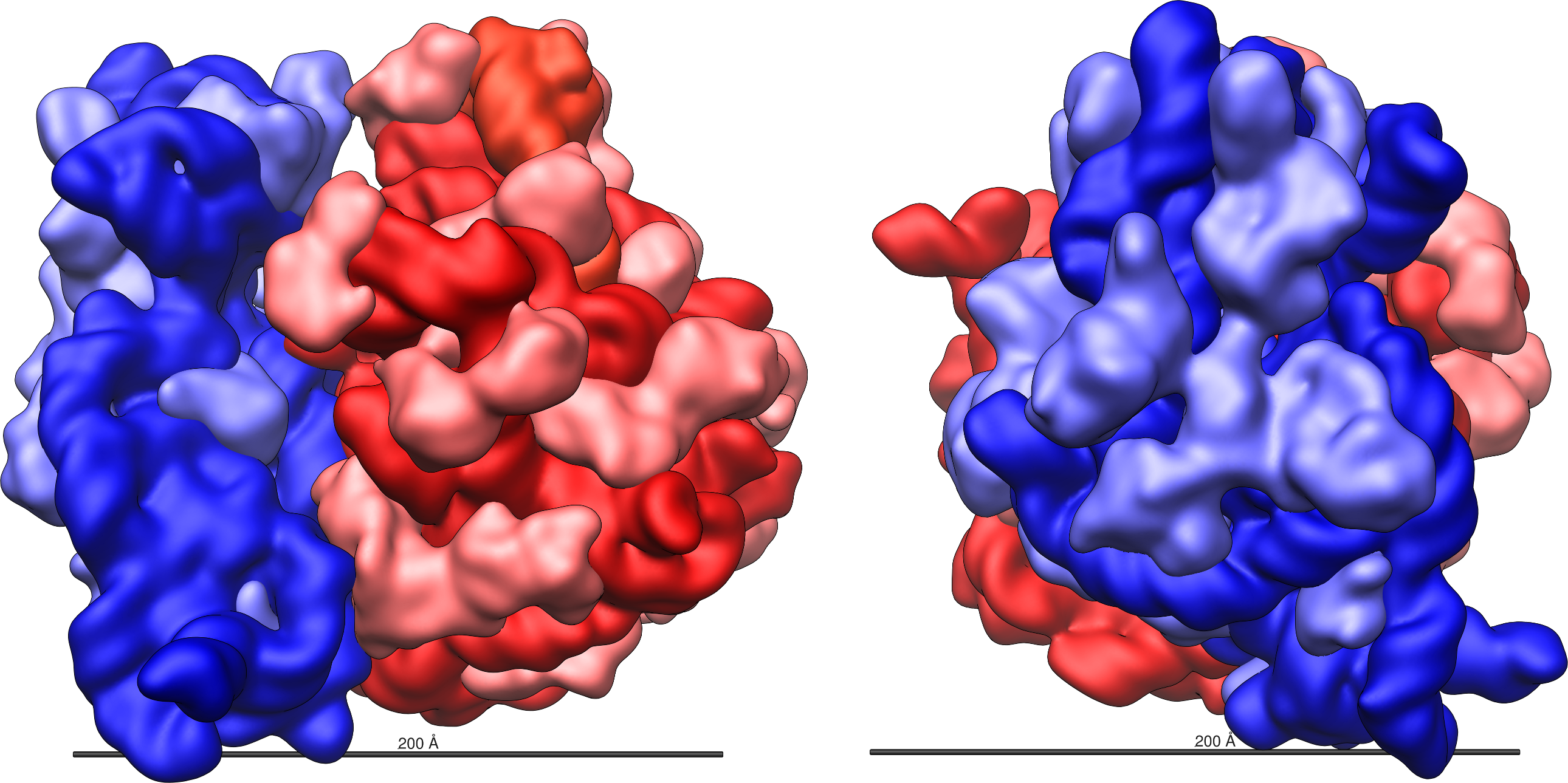
| Region | What it contributes |
|---|---|
| Decoding centre · 30S | Positions mRNA and anticodons; discriminates pairing geometry. |
| Peptidyl-transferase centre · 50S | Aligns the amino-acid-bearing ends of tRNAs for peptide-bond chemistry. |
| A, P and E sites | Accept an incoming aminoacyl-tRNA, hold peptidyl-tRNA, and permit spent tRNA to exit. These are changing binding states, not three static containers. |
| Intersubunit bridges, stalks and exit tunnel | Couple movement and factor binding to the reaction cycle; provide a route for the emerging protein. |
02 · What information is required to make a ribosome?
The instructions are distributed across a genome and an inherited cellular system. There is no single “ribosome blueprint gene.” rRNA genes specify RNA scaffolds directly; ribosomal-protein genes specify amino-acid sequences through translation. Other genes encode RNA polymerase, processing enzymes, assembly factors, tRNAs, aminoacyl-tRNA synthetases, translation factors, and metabolic machinery. Promoters, regulatory RNA structures, and expression levels help coordinate the supply.
| Information layer | Necessary contribution | What should be measured |
|---|---|---|
| RNA sequence and folding | rRNA sequences must form compatible helices, junctions, binding surfaces and functional centres. | Species-specific mature sequences, processing sites and tolerated sequence variants. |
| Protein sequences and interfaces | Ribosomal proteins must bind the appropriate RNA and protein partners without preventing the required motions. | Nonredundant protein sequences; copy number is a separate measurement. |
| Regulation and stoichiometry | Production must match demand and avoid excessive unassembled components. | Promoter behaviour, operon organization, feedback and component abundance. |
| Maturation and chemical environment | Processing, modifications, ions and suitable conditions allow encoded polymers to become active particles. | Enzyme requirements, temperature, ion concentrations, assembly efficiency and activity. |
| Translation correspondence | tRNA identities and amino-acid loading must be compatible with codon recognition. | Charging specificity, anticodons, modifications and release-factor specificity. |
A bacterial set of three mature rRNAs contains roughly 4,600 nucleotides, with exact lengths depending on species and strain. Protein-coding requirements add approximately three DNA nucleotides per amino-acid residue, plus stop and regulatory sequences. One can therefore count sequence length, but that is not yet a calculation of the probability of a functional ribosome. Repeated rRNA operons, alternative functional sequences, shared accessory genes and inherited molecules complicate any total.1
The DNA sequence also does not list the three-dimensional coordinates of every atom. Those coordinates emerge through molecular folding, interactions with other components, solvent and ions. The central origin question is therefore how sequences and a compatible physical system first became coupled, rather than how a genome might literally store an atomic construction drawing.
Back to section guide ↑03 · Atomic precision: A2451, tRNA ends and reaction geometry
Peptide synthesis depends on the arrangement of individual chemical groups over ångström distances: 1 Å is 0.1 nm. The incoming amino acid’s α-amino group must approach the carbonyl carbon of the peptide attached to the P-site tRNA in a productive orientation. Random contact is insufficient. The active site constrains distance, orientation, solvent access and the hydrogen-bond network while allowing the reaction and subsequent release. Measurements comparing ribosomal and solution reactions show that substrate positioning and the reduction of the entropic cost of reaction make a major contribution to catalysis.3
A2451 is an adenosine nucleotide within 23S rRNA, using conventional E. coli numbering. It is not an isolated molecule floating into place for each reaction. Its base, ribose and phosphate backbone occupy a site maintained by the surrounding folded RNA. Its spatial relationship to the two tRNA ends matters, as do the chemistry and orientation of its ribose hydroxyl groups.
A2451, atom by atom
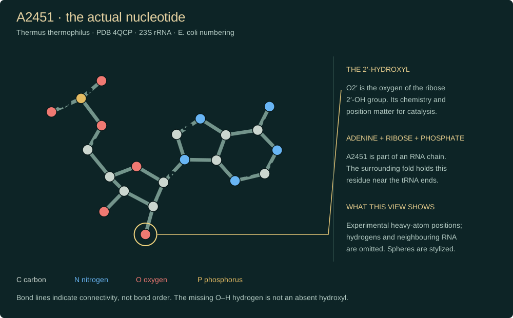
The letter A identifies adenine, while 2451 identifies this residue’s conventional position in the RNA sequence. Adenine is linked to a ribose sugar; the sugar is linked into the phosphodiester backbone. The relevant hydroxyl has an oxygen and a hydrogen attached at the sugar’s 2′ carbon. Losing that oxygen-containing group changes hydrogen-bonding possibilities even when most of the nucleotide remains present. The illustration displays a structure, not an experimental movie or an image of a free adenosine molecule.
Chemical substitutions at A2451 distinguish these requirements. Removing its ribose 2′-OH compromised peptide-transfer activity in a reconstituted system; changing or even removing the adenine base had much smaller effects in those assays when the 2′-OH remained. That finding is more informative than saying that every atom at that site is equally indispensable. In a different set of experiments, substitutions of the A2451 base produced a dominant-lethal phenotype in E. coli but still permitted measurable peptide formation and full-length translation in experimental preparations. Cellular fitness, decoding fidelity and an isolated bond-forming assay are different endpoints.5,7
The terminal adenosine of tRNA, A76, is another critical participant. Replacing the P-site A76 2′-OH with hydrogen or fluorine reduced peptide-bond formation by at least a million-fold under the conditions tested, without abolishing substrate binding. A substrate can therefore sit in the pocket yet fail to react efficiently because a single functional group has changed.4
| Feature | Why location and geometry matter | Evidence boundary |
|---|---|---|
| P-site A76 and A-site amino acid | The reactive groups must be brought together in the appropriate stereochemical relationship. | Binding affinity alone does not establish catalytic competence. |
| A2451 ribose 2′-OH | Contributes to the local functional/structural network around the substrates. | Its role must be separated from early proposals assigning the adenine base a simple general-base mechanism. |
| Ordered waters and nearby RNA | Hydrogen-bond donors and acceptors constrain plausible proton-transfer routes. | A crystallographic contact supports a mechanistic model; it does not directly film proton motion. |
| Ions and surrounding structure | Screen RNA charge and help stabilize folding and interfaces. | Correct bulk composition cannot substitute for a properly assembled local environment. |
Position, orientation and a changing reaction pocket
A 2014 bacterial structural study described a possible proton relay involving P-site A76, A2451 and an ordered water molecule. In that structure the proposed network had donor–acceptor separations below 3.1 Å and successive non-hydrogen-atom angles of about 107–121°. These are observations supporting one model, not universal allowable tolerances for a functioning ribosome. Alternative proton-shuttle descriptions remain part of mechanistic discussion. The general-base role originally assigned to the A2451 adenine should not be presented as settled chemistry.6
Atomic organization extends to decoding. A1492, A1493 and G530 of bacterial 16S rRNA interact with the codon–anticodon helix and change conformation on correct pairing. Recognition of the first two base-pair geometries is especially stringent, whereas the third position permits defined wobble interactions. Atomic-scale precision thus supports both selectivity and a controlled degree of flexibility.2
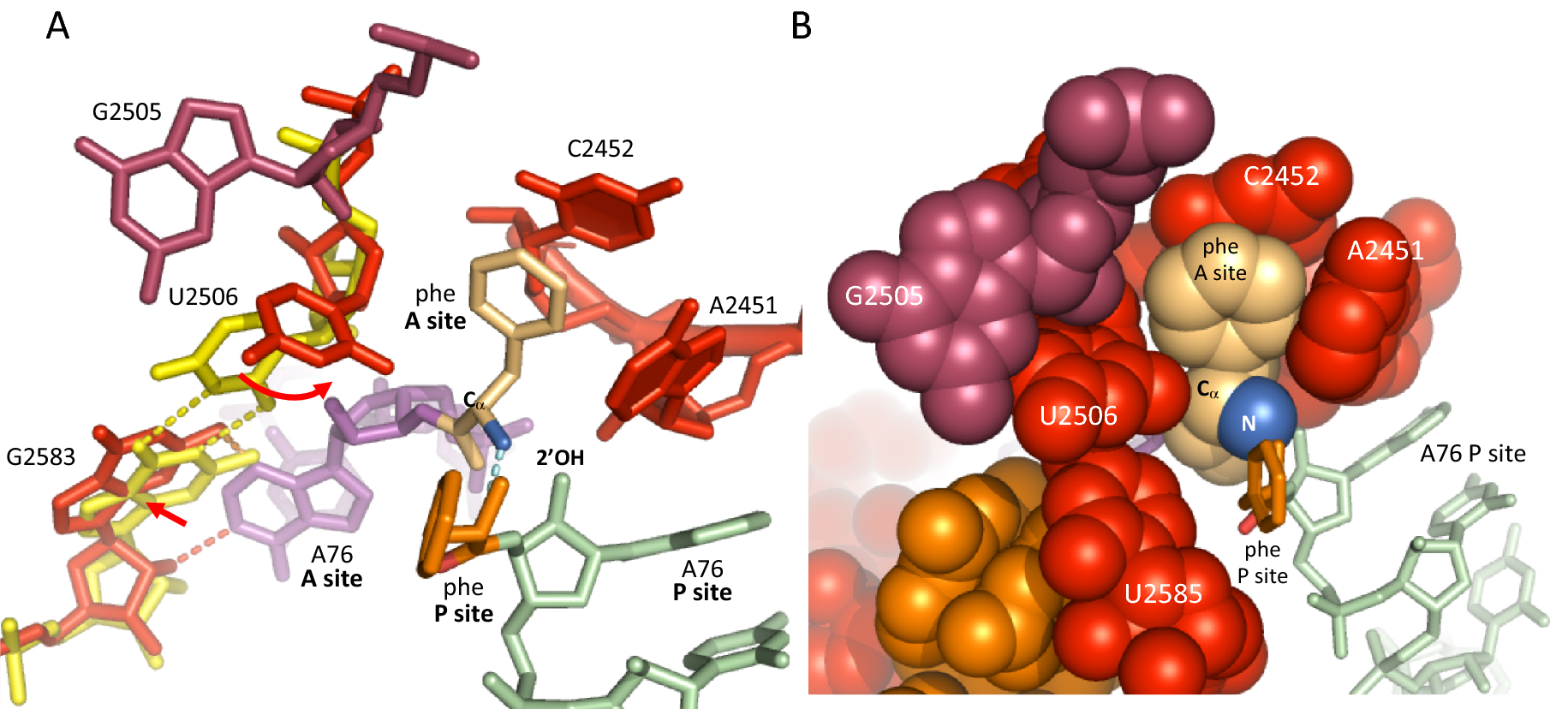
The published comparison above illustrates induced fit: U2506 helps restrict the substrate’s available space as the tRNA CCA end engages the pocket. The authors also propose a thermodynamic explanation for reopening the pocket after peptide transfer. The observed structural arrangements and that proposed energetic explanation should be distinguished.41
Several kinds of precision operate together. Distance permits contact between reacting groups; orientation permits a productive approach to the carbonyl; stereochemistry determines which groups face each other; electrostatics and solvent organization affect charge and proton movement; and timing ensures that a productive arrangement occurs during the correct step. An appropriately placed but chemically altered group can fail, just as a chemically correct group in an inappropriate conformation can fail. These are constraints on an interacting system, not merely a list of ingredients.
Other bacterial sites with demanding chemical or spatial requirements
The following sites show that A2451 is one participant in a distributed network. Residue numbers follow the conventional E. coli scheme; experiments and structures use the bacterial species identified in the cited papers. “Conserved” means retained across the compared sequences; essentiality and the size of a mutation’s effect still require a specified assay.
| Site or group | What must be coordinated | Experimental evidence and limits |
|---|---|---|
| P-site tRNA A76 · ribose 2′-OH | The tRNA-bound ester and incoming amino group must occupy productive relative positions; the neighbouring hydroxyl contributes to the reaction. | Replacing this hydroxyl with H or F produced a ≥10⁶-fold rate loss in the tested peptide-transfer assay, despite preserved binding. This is a reaction-rate effect, not the probability of an origin.4 |
| 23S rRNA A-loop · G2553 | G2553 pairs with C75 at the A-site tRNA end, anchoring the CCA terminus relative to the reaction centre. | In randomized A-loop libraries selected for growth in E. coli, G2553 was retained in every recovered functional sequence, while several neighbouring bases varied.46 |
| 23S rRNA P-loop · G2251 / G2252 | G2251 pairs with tRNA C75 and G2252 with C74, helping position the peptidyl-tRNA terminus. | These guanines were invariant among functional P-loop variants recovered in the same selection. Some surrounding substitutions remained functional; the loop is not uniformly immutable.46 |
| 16S rRNA A1492 / A1493 / G530 | The decoding centre checks the geometry of the codon–anticodon helix, coupling pairing to conformational changes. | Structures of cognate tRNA recognition show a reorganized decoding pocket. Correct pairing geometry is recognized through a network of contacts, with defined flexibility at the wobble position.2 |
| 23S rRNA U2506 / U2585 | The pocket must accommodate substrate entry, peptide transfer and subsequent rearrangement rather than remain permanently locked. | Mutations at conserved inner-shell residues had substrate-dependent effects: puromycin assays and intact aminoacyl-tRNA assays gave different results. Strong conservation does not by itself establish an identical chemical role at every site.42 |
| 23S rRNA A2602 · ribose framework | Peptide release requires a configuration appropriate for water-mediated cleavage of the peptidyl-tRNA ester at termination. | Chemical engineering separated the sugar’s role from the base’s role: an intact ribose was important for efficient release, whereas removing the base could preserve substantial activity. A peptide-release constraint need not be an identical peptide-transfer constraint.44 |
| 23S rRNA A2662 · phosphate oxygen | The sarcin–ricin loop contacts elongation-factor catalytic machinery. The spatial presentation and charge of a nonbridging phosphate oxygen matter. | Atomic substitutions at the two nonbridging oxygens produced different effects on EF-G activation; changing which side was modified changed function. EF4 showed the same trend. This directly tests stereochemical specificity at a backbone group.45 |
| EF-Tu His84 · factor–RNA interface | In a bacterial ribosome–EF-Tu structure, His84 is positioned near the GTP hydrolysis centre through interaction with the A2662 phosphate. | The structure supports a mechanism linking correct decoding to factor activation. Detailed proton-transfer assignments remain mechanistic models; RNA–protein positioning is directly structurally supported.43 |
Why a phosphate’s two oxygen positions can matter
A2662 is especially informative because the experiment changed a backbone chemical group rather than merely exchanging an RNA sequence letter. Replacing either one of its two nonbridging phosphate oxygens with a methyl group produces different stereochemical forms. Those forms did not behave equivalently in ribosome-stimulated GTP hydrolysis. The phosphate’s charge and the presentation of a particular oxygen therefore contribute to activity. This is an experimentally grounded example of “the right group on the right side,” at a site physically separate from the peptide-bond pocket. It does not establish that every phosphate oxygen throughout the ribosome is equally constrained.45
Conservation, compensation and the surrounding fold
An individual contact depends on a larger RNA fold that presents it. Helices and junctions position loops; tRNA pairing helps fix substrate ends; proteins and ions stabilize the assembly; and factor binding changes which states are occupied. A compensatory change can sometimes restore a disrupted interaction, while an apparently minor change at another site can impair folding or slow a crucial transition. The experimental task is to measure the combined effects, rather than assume that every site varies independently.
In the genetic selection study, the five-position A-loop library contained 1,024 possible sequences and yielded nine functional sequences, including the natural one. The corresponding P-loop selection recovered twelve variants. These are useful local measurements of constraint in an already functioning E. coli system. They are not a survey of all possible ribosomes or a reconstruction of an ancestral population. Recovery also depends on the growth conditions and the selection’s sensitivity.46
Related library discussions: Cells and proteins need to be organized on an atomic scale · Fine-tuning of the dihedral angles in the reaction center of enzymes. The experimental distinctions above identify which precision claims are directly supported.
What this establishes about chance, origins and design
A design interpretation can draw attention to coordinated interfaces, chemically specific groups, staged operation and the dependence of each step on the others. Those observations motivate an argument about organized function. They do not independently measure an overall probability of a ribosome arising, or identify the historical process that produced it. The modern ribosome’s constrained sites are evidence about its present mechanism; their origin requires additional historical and experimental evidence. See origins and proposed intermediates and the design discussion for the competing interpretations.
It would be misleading to multiply the inverse of every measured rate effect, or the odds of every exact modern nucleotide, and call the result “the chance of a ribosome.” Reaction-rate ratios are not event probabilities. Sites interact, alternative functional sequences exist, molecular self-assembly is constrained by chemistry, and evolutionary models include inherited variation and selection. At the same time, invoking selection does not by itself demonstrate a complete route from prebiotic chemistry to a translating system. A defensible claim of extreme improbability needs a defined random process, a specified functional target, plausible resources and timescales, and evidence about the accessible alternatives.
How to read atomic pictures and mutation results
| Question | What to check |
|---|---|
| Is this an experimental structure? | Identify the organism, PDB accession, residue numbering and trapped state. A crystal or cryo-EM model is an interpretation of measurements, not a direct photograph of every atom. |
| Are substrates natural? | Stable substrate analogues can trap otherwise short-lived states. Their geometry is informative, but they need not reproduce every property of a reacting substrate. |
| What does a drawn line mean? | A covalent bond, close contact and proposed hydrogen bond are different claims. Heavy-atom structures often do not locate hydrogens directly; proton paths require additional evidence. |
| Does a distance define a tolerance? | A measured separation in one structure is not an experimentally determined permissible range. Neither map resolution nor coordinate decimal precision is a biological manufacturing tolerance. |
| What was actually tested? | Growth, assembly yield, binding, fidelity, peptide-transfer rate and peptide release answer different questions. A mutant may pass one assay and fail another. |
| Can the origin probability be calculated? | Only after specifying the process and the distribution of functional alternatives. A lethal mutation in a modern cell does not show that every historical intermediate was impossible. |
04 · Building the machine: transcription, folding and maturation
Bacterial ribosomes are assembled in the cytoplasm, with folding and protein binding beginning while rRNA is being transcribed. The process combines intrinsic molecular affinity with assisted maturation. It is hierarchical, cooperative and partly parallel: some interactions create binding sites for later components, but there is not one universal assembly sequence followed by every particle.35
Produce compatible components
RNA polymerase transcribes rRNA precursors and ribosomal-protein mRNAs. Existing ribosomes translate the protein messages. Appropriate nucleotides, amino acids, charged tRNAs and energy supplies must be available.
Process and fold the RNA
Precursor RNA is cut and trimmed to mature ends. Folding produces local helices and longer-range contacts; modification enzymes alter selected nucleotides. Processing routes and enzyme requirements vary between bacteria.
Bind proteins and remodel intermediates
Early proteins stabilize RNA folds; later proteins bind newly formed interfaces. RNA helicases, GTPases and other assembly factors can help overcome kinetic traps and coordinate maturation. These temporary factors are not all permanent ribosome parts.
Complete functional centres
The decoding region, subunit interface and peptidyl-transferase centre must reach compatible mature configurations. Large-subunit cryo-EM intermediates reveal late formation of important functional regions.
Release or rework the particle
Mature subunits enter translation; defective intermediates may be remodelled or degraded. Assembly-factor release and factor reuse link one construction cycle to the next.
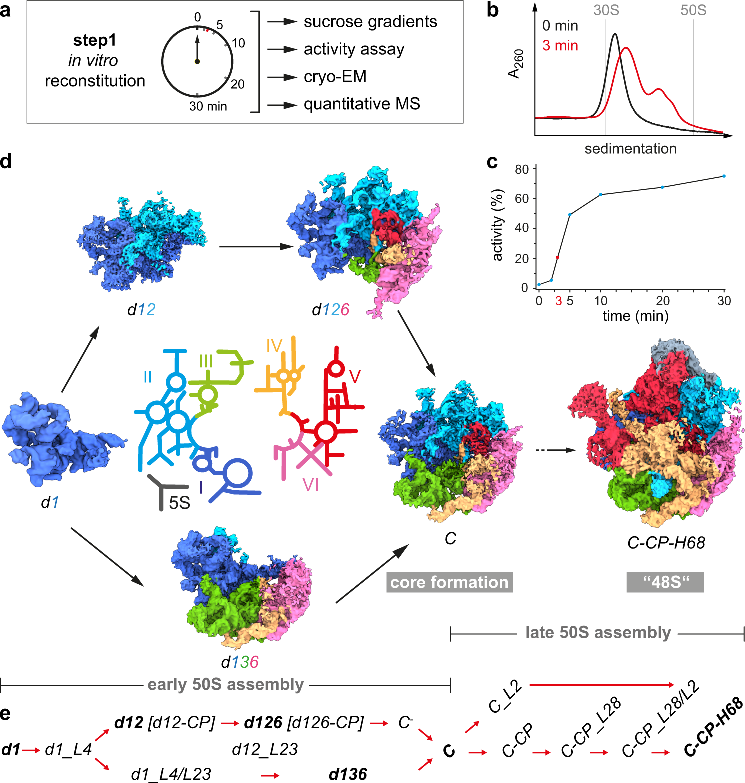
The reconstitution images above resolve intermediate RNA domains and protein occupancy into an experimentally constrained assembly pathway.9
“Self-assembly” means that molecular interactions contribute the ordering; it does not mean that an arbitrary mixture of small molecules spontaneously makes a ribosome. Conversely, the need for assistance in living cells does not mean that purified, already synthesized components lack assembly capacity. Classic experiments reconstructed bacterial large subunits from supplied RNA and protein components at high temperatures. A mechanistic account must specify the starting materials and experimental conditions.34
Bacterial quality control includes active remodelling. A 2023 KsgA study found that the methyltransferase recognizes inactive small-subunit particles and partially disassembles them, opening a route toward productive reassembly. The result supports an assembly-proofreading role; it is not evidence that every incorrect nucleotide can be repaired in place.10
The adaptors must also be manufactured
An active ribosome is insufficient if its tRNA supply is immature. In the classic bacterial RNase P system, a catalytic RNA and a protein partner process the 5′ ends of precursor tRNAs. Experiments showed that the RNA component can catalyse cleavage under appropriate laboratory conditions; the normal cellular enzyme’s protein partner contributes to efficient function.37
The tRNA acceptor end must also present the CCA terminus, ending in A76, used for amino-acid attachment and ribosomal interactions. Bacterial CCA-end maturation has more than one enzymatic arrangement. Structural work on an Aquifex aeolicus enzyme bound to tRNA lacking its final adenosine and an incoming ATP analogue shows how this endpoint is constructed with molecular specificity. Correct RNA ends, folding, selected modifications and charging are therefore part of the translation system’s manufacturing requirements, even though they are not components of the ribosome particle itself.36
Back to section guide ↑05 · Signals that regulate production and operation
“Signaling” operates on several scales. A diffusible regulator can change gene expression across the cell; a protein can repress its own operon; and a local structural change can alter the next reaction on a single ribosome. These are distinct physical mechanisms. None requires a separate electronic control circuit or a conscious decision-maker inside the cell.
| Input or state | Bacterial mechanism | Functional outcome |
|---|---|---|
| Amino-acid shortage | In E. coli, uncharged tRNA at an appropriate stalled ribosome activates RelA and production of (p)ppGpp. | Connects translation demand to the stringent response.12 |
| (p)ppGpp and transcription conditions | In the studied E. coli system, DksA and ppGpp alter RNA-polymerase initiation at rRNA promoters. | Reduces investment in ribosome production during shortage.11 |
| Excess unassembled ribosomal protein | Some ribosomal proteins bind regulatory sites on their own operon’s mRNA; L1 provides a classic example. | Negative feedback helps balance protein production with assembly demand.13 |
| Correct codon–anticodon pairing | Conformational coupling accelerates EF-Tu GTPase activation and tRNA accommodation. | Links recognition to commitment through kinetic selection.8 |
| An emerging targeting sequence | Bacterial SRP and its receptor FtsY coordinate delivery toward membrane translocation machinery. | Connects protein synthesis to localization.14 |
| Prolonged stalling and collision | In E. coli, SmrB recognizes a collided-ribosome arrangement and cleaves the message. | Converts a stalled queue into substrates for rescue.19 |
This table describes experimentally studied examples, not a claim that every named factor has the same role in Aquifex. Bacterial lineages differ in their regulatory and rescue repertoires. The atlas’s accession list is the starting point for checking which components are present; functional equivalence requires additional evidence.
During elongation, long-range coupling coordinates a decoding event in the small subunit with events at the factor-binding regions and the tRNA ends in the large subunit. tRNA deformation, subunit rotation, head movement and factor binding are parts of the physical cycle. Descriptions such as “communication” are useful shorthand for these changes in molecular interaction and reaction probability.8
Back to section guide ↑06 · Operation, energy, timing and the finished protein
Initiation establishes the reading frame. The small subunit, initiation factors and initiator tRNA organize an mRNA start region; the large subunit joins to establish an elongation-ready complex. Many bacterial messages use a Shine–Dalgarno interaction, but bacterial initiation is diverse and includes leaderless messages. AUG commonly specifies the start, and alternative bacterial start codons exist. Initiator methionine is generally formylated in bacterial initiation.
Elongation coordinates selection, chemistry and movement. EF-Tu–GTP delivers aminoacyl-tRNA. Correct recognition favours GTPase activation; accommodation brings the acceptor end into the large-subunit active site. Peptidyl transfer moves the growing chain onto the incoming tRNA. EF-G and GTP support translocation so the message advances by one codon. EF-Ts helps regenerate EF-Tu’s active nucleotide-bound state. Purified-component translation experiments demonstrate how these supplied factors can support protein synthesis outside a living cell.8,26
Termination is a chemically different use of the active site. Bacterial RF1 and RF2 recognize stop signals and promote hydrolysis of peptidyl-tRNA, releasing the protein. Stop recognition must be distinguished from the later splitting and reuse of the ribosome. Readthrough or frameshifting can occur as regulated exceptions or errors; they should not be confused with ordinary triplet elongation.
Useful speed must be balanced against accuracy. Rejecting a near-cognate tRNA costs time and can waste energy, whereas accepting it risks a defective product. A persistent pause can obstruct trailing ribosomes, but some pauses assist regulation or folding. There is no single error rate or elongation speed valid for all bacteria, sequences, temperatures and growth conditions. Nor does a productive peptide-bond event prove that the full translation cycle will run efficiently.
The emerging chain is not automatically a working protein. Sequence, solvent and the cellular environment determine folding; chaperones can limit aggregation or aid maturation. A membrane protein may require SRP-directed targeting and insertion, while other proteins fold in the cytoplasm or require cofactors. A complete origin account must connect useful protein production to these downstream requirements, rather than stop at the formation of an arbitrary peptide.14
Back to section guide ↑07 · The genetic code: the correspondence that translation implements
The genetic code is the mapping from mRNA triplets to amino acids or termination signals. It is different from a particular gene sequence. In the standard code, 64 triplets comprise 61 amino-acid codons and three stop codons. Multiple codons can specify one amino acid. Such degeneracy does not make all substitutions harmless, and a codon’s effect also depends on reading frame and context.
The correspondence is physically implemented by tRNAs, their recognition by synthetases or other aminoacylation pathways, and the ribosome’s decoding machinery. The ribosome mostly evaluates the codon–anticodon interaction; it does not independently verify the identity of every attached amino acid. Correctly pairing but mischarged tRNAs can therefore deliver the wrong residue. tRNA identity elements extend beyond the anticodon, including sites in the acceptor region, so the charging correspondence cannot be reduced to anticodon pairing alone.32,15
Open the standard genetic-code table · all 64 RNA codons
Triplets are written 5′ → 3′. This is the standard mapping, not a complete map of bacterial initiation rules or exceptional recoding events. Met = methionine; Stop = termination.
| First two bases (N = third) | All four third-base possibilities |
|---|---|
| UUN | UUU Phe · UUC Phe · UUA Leu · UUG Leu |
| UCN | UCU Ser · UCC Ser · UCA Ser · UCG Ser |
| UAN | UAU Tyr · UAC Tyr · UAA Stop · UAG Stop |
| UGN | UGU Cys · UGC Cys · UGA Stop · UGG Trp |
| CUN | CUU Leu · CUC Leu · CUA Leu · CUG Leu |
| CCN | CCU Pro · CCC Pro · CCA Pro · CCG Pro |
| CAN | CAU His · CAC His · CAA Gln · CAG Gln |
| CGN | CGU Arg · CGC Arg · CGA Arg · CGG Arg |
| AUN | AUU Ile · AUC Ile · AUA Ile · AUG Met |
| ACN | ACU Thr · ACC Thr · ACA Thr · ACG Thr |
| AAN | AAU Asn · AAC Asn · AAA Lys · AAG Lys |
| AGN | AGU Ser · AGC Ser · AGA Arg · AGG Arg |
| GUN | GUU Val · GUC Val · GUA Val · GUG Val |
| GCN | GCU Ala · GCC Ala · GCA Ala · GCG Ala |
| GAN | GAU Asp · GAC Asp · GAA Glu · GAG Glu |
| GGN | GGU Gly · GGC Gly · GGA Gly · GGG Gly |
The code contains an error-buffering pattern: many neighbouring codons encode the same or chemically similar amino acids. Computational comparisons find substantial error minimization under defined cost functions and random-code ensembles. A ranking such as “one in a million” is conditional on those choices; it is not the probability that the code originated naturally, nor a posterior probability for design.31
Origins proposals include chemical affinities between RNA and amino acids, expansion associated with amino-acid biosynthesis, selection for error tolerance, and historical fixation once changing assignments became costly. Structural analyses report enrichment of some cognate triplets near amino-acid residues, giving limited support to stereochemical ideas. These observations do not derive the complete code or demonstrate a self-sustaining ancestral translation system.30
The difficult transition is from chemistry that sometimes joins amino acids to an inheritable correspondence in which message sequence reliably controls useful peptide sequence. A candidate history must connect adaptor formation, charging specificity, readable templates, catalytic activity, and a benefit that can be retained. Explaining one assignment or one chemical step does not by itself explain the whole mapping.
Back to section guide ↑08 · Error prevention, detection and rescue
Quality control is distributed across the supply chain. It is misleading to give one fixed number of “ribosomal repair systems”: lists often mix tRNA maturation, charging, decoding, rescue, protein degradation and rRNA turnover. These mechanisms address different faults and are not interchangeable. The following map keeps those distinctions visible.
| Checkpoint | Fault being addressed | Bacterial response and limit |
|---|---|---|
| RNA maturation and assembly | Misprocessed RNA or improperly assembled subunits. | Processing and assembly factors help produce functional particles. KsgA can remodel inactive small-subunit states; YbeY participates in 16S maturation and defective-ribosome quality control.10,22 |
| tRNA charging | A synthetase activates or transfers an inappropriate amino acid. | Substrate discrimination, pre-transfer editing, post-transfer hydrolysis and separate editing factors can improve fidelity. Different synthetases use different combinations; they do not all share one editing mechanism.15 |
| Initial selection and accommodation | A near-cognate tRNA encounters a codon. | EF-Tu-linked selection and later proofreading favour correct substrates before bond formation. Some errors escape; this is kinetic discrimination, not perfect base-by-base certainty.8 |
| After peptide formation | A miscoding event leaves a mismatched codon–anticodon arrangement. | Defined bacterial assays show altered subsequent selection and premature termination. The wrong amino acid is not simply excised and replaced within the completed chain.16 |
| A truncated message | A ribosome reaches an mRNA end without a usable stop signal. | tmRNA–SmpB can rescue the complex and append a degradation tag to the incomplete product.17 |
| A blocked ribosome queue | A trailing ribosome catches a persistently stalled one. | SmrB-dependent message cleavage in E. coli and MutS2-associated responses in B. subtilis connect collision sensing to downstream quality control.19,20 |
| A defective ribosome | A functional centre or RNA component cannot support normal activity. | Selective RNA degradation can remove defective particles; salvage is different from correction of the original sequence.22,23 |
tRNA editing is upstream of the ribosome. For example, an editing site can hydrolyse a wrongly attached amino acid, freeing the tRNA for another charging attempt. This is genuine chemical correction at the charging stage. It differs from rejecting a mismatched anticodon or destroying a defective protein. Amino-acid chirality, tRNA modifications and the chemical similarity of substrates also affect discrimination; the ribosome is not an infallible final inspector for all upstream mistakes.
Rescue protects both the ribosome pool and proteome. tmRNA combines a tRNA-like function with a short coding template. Together with SmpB it allows translation to switch away from a defective message, add a tag, terminate, and free the machinery; the tag helps target the incomplete product for degradation. This is a coordinated response to a stalled complex, not ordinary translation of an intact message.17
Alternative rescue systems are distributed unevenly across bacteria. In E. coli, ArfA senses an empty downstream mRNA channel and recruits RF2 for stop-independent release. Structural work connects the sensing state to activation of RF2’s catalytic configuration. ArfA-mediated release should not be described as automatically attaching the tmRNA degradation tag; its immediate outcome is peptide release and rescue.18
Collisions provide a geometrical signal larger than a single stalled ribosome. SmrB in E. coli recognizes a composite arrangement and cuts mRNA upstream of the stalled particle, enabling rescue of upstream ribosomes. In B. subtilis, MutS2 links collision recognition to splitting-associated quality control and nascent-chain tagging. These are experimentally distinguished bacterial solutions, not a universal checklist that every minimal ribosome must contain.19,20
Back to section guide ↑09 · Repair, recycling, degradation and storage
| Process | What happens | What it does not establish |
|---|---|---|
| Normal recycling | RRF and EF-G split a post-termination complex; IF3 helps stabilize separation so components can be reused.21 | The RNA and protein subunits need not be destroyed after every protein is made. |
| Assembly reworking | An inactive assembly intermediate can be remodelled and given another route to maturation.10 | There is no general mechanism known to rewrite every damaged rRNA nucleotide back to its original sequence. |
| Specific rRNA repair | PrfH-associated rescue and the RNA ligase RtcB2 can restore ribosomes with defined decoding-centre RNA cleavage.38 | Repair of a particular RNA break is not correction of arbitrary substitutions or all forms of damage. |
| Protein replacement | Exchangeable bacterial ribosomal proteins can be replaced, and damaged preparations can regain translation activity.40 | This does not mean all deeply embedded ribosomal proteins can be extracted and replaced in the same way. |
| Defective-ribosome turnover | YbeY and RNase R can cooperate in selective removal of defective bacterial ribosomes.22 | Destroying a defective particle is not the same as restoring that particle. |
| RNA breakdown and salvage | Endonucleolytic cleavage and exonucleolytic processing contribute to rRNA degradation during quality control or starvation.23 | Bacteria do not use a nucleolus-to-lysosome ribophagy pathway as their default explanation. |
| Hibernation | Some bacteria protect inactive ribosomes using hibernation factors; 100S particles are dimers of 70S ribosomes.24 | The dimer is not a bigger translating ribosome and is not universal across bacteria. |
Direct repair of bacterial rRNA is experimentally supported. Tian and colleagues studied a system in which PrfH recognizes a specific cleavage in the decoding centre and promotes release of the attached peptide; RtcB2 then ligates the broken RNA. Their biochemical, structural and cell-based experiments distinguish recognition of the damaged state from ordinary stop-codon termination. The pathway is not present as a functional universal module in every strain: the standard E. coli K-12 lineage has a disrupted rtcB2–prfH region.38
A 2026 follow-up on colicin E3 damage found that RtcB2 alone could repair cleaved 30S and 70S particles under tested conditions, while PrfH increased ligation efficiency for ribosomes engaged in translation. This qualifies an overly rigid account in which repair must always await complete dismantling. The relevant lesson is sequential, state-dependent coordination, with the substrate and experimental conditions stated.39
Protein exchange offers another restorative route. Pulk and colleagues restored translational activity of damaged bacterial ribosomes by supplying ribosomal proteins and detected protein exchange in stationary cultures. The exchangeable group was associated with relatively accessible, loosely bound proteins. These results support repair by replacement for a subset of components; they do not demonstrate a universal extraction mechanism for every ribosomal protein.40
“Repair” should be used for a demonstrated restorative process. Reworking an assembly intermediate, sealing a specific RNA break, replacing a damaged protein, recycling after termination, and degrading an incomplete product are different outcomes. A bacterial account can include all of them without borrowing mechanisms from other domains of life.
Maintenance also consumes resources. Degraded RNA can supply recoverable building blocks, but a cell still needs synthetic pathways to rebuild components and energy to operate the system. Hibernation temporarily preserves an investment; it does not make the ribosome independent of metabolism. In E. coli, RMF and a short HPF cooperate in 100S formation, whereas long-form HPF supports a different dimerization architecture in B. subtilis.24
Back to section guide ↑10 · What would a minimal bacterial ribosome have to be?
A meaningful minimum is a minimum for a stated task in stated conditions. Counting components without defining activity, accuracy, temperature, nutrient supply and lifetime hides the central issue. A catalytic fragment, an externally supported translation system and a self-maintaining bacterium answer different questions.
| Target | Required demonstration | Status and limitation |
|---|---|---|
| Peptide-bond catalyst | Promotes a specified bond-forming reaction using supplied substrates. | Engineered protoribosome-like RNA constructs show limited bond formation. This does not demonstrate codon reading, processive translation or reproduction.33 |
| Minimal translating particle | Reads an mRNA in frame and repeatedly makes a defined product using supplied tRNAs and factors. | Requires coordinated decoding, catalysis, movement and release. Component dispensability depends on assay conditions; a peptide-formation assay alone is insufficient. |
| Minimal translation system | Maintains charging, initiation, elongation, termination and reuse with enough fidelity and yield. | Purified systems demonstrate activity with externally supplied mature components and energy. They are not automatically self-regenerating.26 |
| Ribosome-producing system | Makes functional new subunits through transcription, protein synthesis, processing and assembly. | A 2025 E. coli-extract system achieved this in one reaction space using supplied genes and biological machinery.27 |
| Self-maintaining bacterial cell | Continually replaces the translation system while supporting metabolism, genome maintenance, a membrane and division. | The atlas’s 912-protein reconstruction is a working inventory, not an experimentally demonstrated viable minimum or the first cell. |
No universal “53 proteins plus three RNAs” threshold is established. Shoji and colleagues reported that, combining their results with earlier work, 22 of 54 E. coli ribosomal-protein genes could be deleted individually under tested conditions. This does not establish a viable ribosome missing all 22 at once: multiple deletions can interact, growth defects matter, and the cell still supplies assembly factors and other support. It also does not turn the remaining 32 into a proven universal minimum.25
A plausible minimal translating system would still need: a decoding scaffold; a bond-forming centre with productive substrate geometry; adaptors and a compatible amino-acid assignment system; access to charged substrates and energy; a means to establish a reading frame, advance and terminate; and sufficient component stability or replacement. These are functional requirements, not proof that today’s exact proteins, RNA lengths or factor inventory had to exist at the beginning.
The 2025 biogenesis experiment is an important advance: it combined synthesis and assembly of functional new small and large subunits in an optimized E. coli extract. Its reaction contained a soluble cellular proteome and started from supplied genetic and translation resources. It did not generate a translation system from prebiotic feedstocks or establish indefinite reproduction of an autonomous cell. Both the achievement and the boundary belong in an accurate account.27
What a convincing minimum experiment should report
- Exactly which RNA, protein and accessory factors are present, absent or supplied externally.
- Successful production of diverse, full-length proteins—not just one bond or one permissive reporter.
- Accuracy, frameshifting, readthrough, speed, yield and stability over repeated cycles.
- Combined-deletion tests, rescue experiments and controls for contamination by intact ribosomes.
- Whether newly made ribosomes contribute to making subsequent ribosomes, with dilution and replenishment conditions stated.
- For a cellular claim: growth and reproduction in a chemically defined environment, with all environmental dependencies explicit.
11 · Origins: what is known, inferred and still missing
The bacterial ribosome is ancient. Shared structural features and an RNA-centred catalytic core motivate proposals that simpler RNA–peptide systems preceded modern translation. Comparative structural reconstructions infer growth by accretion of RNA elements, linking proposed earlier stages with gains in function. Such models use present-day structures to reconstruct history; they do not directly observe the proposed ancestors or determine one uniquely established path.28
Three distinctions prevent an origins discussion from becoming misleading. First, the ancestor shared by extant cells is not necessarily the first life. Second, requirements for modern bacterial growth do not prove that every modern accessory pathway existed before any heritable selection. Third, a demonstration of molecular self-assembly from purified biological components is not a demonstration of their prebiotic synthesis.
| Origin problem | Why it is difficult | What partial evidence does—and does not—show |
|---|---|---|
| Building and preserving components | Useful RNA and peptide sequences must be produced, persist and reach compatible concentrations. | A mechanism must address chemistry, degradation and environmental compatibility, not assume purified laboratory supplies. |
| From catalysis to coding | Joining amino acids is different from choosing their sequence from a template. | Protoribosome-like constructs address aspects of catalysis, leaving adaptor assignments and processive decoding to explain.33 |
| Bootstrapping | Modern ribosomes make proteins needed for modern ribosome production. | A historical model needs an earlier, simpler source of useful catalysts or a coevolving RNA–peptide network; present-day circular dependence alone does not prove impossibility. |
| Heritable improvement | Any proposed intermediate must have a selectable benefit associated with inheritable variation. | RNA-enzyme replication experiments demonstrate restricted heredity without proteins, but use designed molecules and supplied substrates—not a complete prebiotic cell.29 |
| Fidelity and error burden | Poor copying and translation can prevent useful functions from persisting. | The required fidelity depends on sequence length, redundancy, fitness effects and replication dynamics; there is no universal modern error threshold for all ancestors. |
| Coordinated chemistry | Charging, RNA folding, catalysis, energy use and compartmentalization must work together. | Compatibility must be demonstrated across a coupled system, not inferred solely by combining isolated successes. |
| Explaining the modern code | Assignments must become stable enough to support useful proteins yet allow earlier expansion or refinement. | Stereochemical and error-minimization evidence constrains proposals without supplying a complete historical reconstruction.30,31 |
The fact that proteins are needed by modern translation creates a substantive bootstrap question. It does not logically exclude selection before DNA-and-protein cells: any system with replication, heritable variation and differential persistence can undergo selection. RNA cross-replication experiments make that distinction concrete, while leaving substantial challenges in prebiotic plausibility and open-ended reproduction.29
A responsible comparison asks which model explains the transition with the fewest unsupported assumptions and the best experimental or comparative support. Failure to reconstruct every step is an open research problem; it is not itself a measured probability of impossibility. Equally, listing plausible ingredients or isolated reactions does not amount to an experimentally demonstrated origin of translation.
Back to section guide ↑12 · Hallmarks of design: an explicit argument to examine
FUNCTIONAL ORGANIZATION · AUTHOR’S FRAMEWORK
Hallmarks of design
Bacterial ribosome illustrates the following organizational features in the supplied papers. These descriptions identify biological functions; interpreting their origin as design is the author’s comparative argument.H1, H2
Adaptors bridge anticodon recognition and amino-acid delivery; charging establishes amino-acid identity.
The genome specifies components of the translation machinery that expresses that genome.
Assembly surveillance, selection, proofreading and rescue act at different stages.
Information, energy, substrates and ångström-scale reaction geometry operate together.
Origin hurdles and abductive assessment · Assessment method · All 40 hallmarks. Biological evidence: the mechanism, accession records and scientific references on this page.
The features that motivate a design interpretation are concrete: sequence-dependent production, a distributed codon-to-amino-acid correspondence, complementary interfaces, atomic-scale reaction geometry, energy-coupled checkpoints, component manufacturing, feedback, fault detection and reuse. In engineered systems, combining these capabilities commonly reflects planning. The ribosome therefore supplies a serious case for examining a design analogy rather than merely noting that biology is complicated.
| Observed feature | Design-oriented interpretation | Question an explanation must answer |
|---|---|---|
| Coded sequence control | A material message is interpreted through adaptors and a mapping, analogous to a programmed production system. | How did an inheritable, useful correspondence arise and become stabilized? |
| Atomic coordination | Catalytic groups, substrate ends and waters cooperate at the scale of individual functional groups. | Which geometries are necessary, which variations are tolerated, and can simpler productive arrangements be demonstrated? |
| Interdependent subsystems | The machine, its components and their manufacturing processes support one another. | Can a proposed historical route identify useful, viable intermediates rather than only an endpoint? |
| Monitoring and recovery | Fault-specific responses preserve productive translation and recover resources. | Which mechanisms are required under which conditions, and which are later improvements? |
| Integrated performance | Speed, fidelity, energy costs and component supply are coordinated. | Do competing accounts explain the measured tradeoffs and constraints, not simply the presence of many parts? |
A clear design argument can be stated as an inference: intelligence is known to produce coordinated, function-directed systems; translation exhibits several analogous features; therefore intelligent causation is proposed as an explanation. Its strength depends on how diagnostic those features are after comparison with chemical self-organization, selection, historical constraints and available experiments. The observed mechanisms are empirical findings; the conclusion about their ultimate cause is an additional interpretive step.
The inference is not strengthened by describing A2451 as an all-or-nothing switch when experiments reveal more nuanced effects, treating every modern factor as primordial, or using the probability of one exact modern sequence as the probability of all possible functional histories. Those shortcuts can be tested and can fail even if the broader design question remains worth discussing.
The central explanatory demand is the origin of an integrated, heritable translation system that can make useful products and help maintain itself. Its precision and interdependence make that demand substantial. They justify detailed investigation; they do not remove the need to distinguish observation, mechanistic model, historical inference and philosophical interpretation.
Back to section guide ↑13 · Terms and practical questions
| Term | Meaning in this section |
|---|---|
| Ribozyme | RNA contributing catalytic function; the ribosomal peptide-transfer centre is RNA-centred. |
| Cognate / near-cognate | An appropriately matching substrate / a close alternative that can sometimes be misaccepted. |
| Accommodation | Movement of an incoming tRNA’s amino-acid-bearing end into the bond-forming site after delivery. |
| Allostery | A change at one region alters behaviour at another through coupled structure and interactions. |
| Kinetic proofreading | Energy-linked sequential selection that can improve discrimination beyond a single binding step. |
| Reconstitution | Rebuilding an active system using supplied, defined or partially defined components. |
| Essentiality | Requirement for an experimentally specified organism and condition; not a timeless property independent of context. |
Does the ribosome correct a wrong amino acid already inside a protein?
It has no general DNA-polymerase-like backspace that removes one wrong internal amino acid and resumes the original chain. Prevention, rejection, abortive termination, rescue and degradation are different quality-control outcomes.
Does atomic precision imply that only one sequence can work?
No. Some chemical groups are strongly constrained, while many sequence changes retain function or change performance. The task is to measure those tolerances rather than infer that all positions are equally fixed.
Do all bacteria use every pathway described here?
No. Examples are labelled by organism. A claim about the proposed Aquifex cell needs accession-resolved and organism-specific support. Do not transfer the complete E. coli regulatory network into that model without checking.
Why do antibiotics matter to this explanation?
Antibiotics can perturb particular binding sites or conformational steps, helping experimentally distinguish functions. Ogle and colleagues showed that paromomycin can favour conformations that permit near-cognate tRNA binding. Such perturbations test molecular specificity; they are not direct tests of an origin scenario.2
Represented in the 912-protein inventory
53 selected protein entries. The ribosome also contains essential ribosomal RNA. RNA molecules are outside this protein-only inventory.
| Gene / locus | Protein entry | Source record |
|---|---|---|
| rpsJaq_008 | Small ribosomal subunit protein uS10 (30S ribosomal protein S10) | O66430 ↗ |
| rplCaq_009 | Large ribosomal subunit protein uL3 (50S ribosomal protein L3) | O66431 ↗ |
| rplDaq_011 | Large ribosomal subunit protein uL4 (50S ribosomal protein L4) | O66432 ↗ |
| rplWaq_012 | Large ribosomal subunit protein uL23 (50S ribosomal protein L23) | O66433 ↗ |
| rplBaq_013 | Large ribosomal subunit protein uL2 (50S ribosomal protein L2) | O66434 ↗ |
| rpsSaq_015 | Small ribosomal subunit protein uS19 (30S ribosomal protein S19) | O66435 ↗ |
| rplVaq_016 | Large ribosomal subunit protein uL22 (50S ribosomal protein L22) | O66436 ↗ |
| rpsCaq_017 | Small ribosomal subunit protein uS3 (30S ribosomal protein S3) | O66437 ↗ |
| rplPaq_018 | Large ribosomal subunit protein uL16 (50S ribosomal protein L16) | O66438 ↗ |
| rpmCaq_019 | Large ribosomal subunit protein uL29 (50S ribosomal protein L29) | P56613 ↗ |
| rpsQaq_020 | Small ribosomal subunit protein uS17 (30S ribosomal protein S17) | O66439 ↗ |
| rpsFaq_063 | Small ribosomal subunit protein bS6 (30S ribosomal protein S6) | O66474 ↗ |
| rpsRaq_064.1 | Small ribosomal subunit protein bS18 (30S ribosomal protein S18) | O66476 ↗ |
| rplQaq_069 | Large ribosomal subunit protein bL17 (50S ribosomal protein L17) | O66482 ↗ |
| rpsDaq_072 | Small ribosomal subunit protein uS4 (30S ribosomal protein S4) | O66484 ↗ |
| rpsKaq_073 | Small ribosomal subunit protein uS11 (30S ribosomal protein S11) | O66485 ↗ |
| rpsMaq_074 | Small ribosomal subunit protein uS13 (30S ribosomal protein S13) | O66486 ↗ |
| rpmJaq_075 | Large ribosomal subunit protein bL36 (50S ribosomal protein L36) | O66487 ↗ |
| rpmBaq_080 | Large ribosomal subunit protein bL28 (50S ribosomal protein L28) | O66492 ↗ |
| rpmFaq_1102 | Large ribosomal subunit protein bL32 (50S ribosomal protein L32) | O67187 ↗ |
| rpsPaq_123 | Small ribosomal subunit protein bS16 (30S ribosomal protein S16) | O66523 ↗ |
| rpsAaq_1485 | Small ribosomal subunit protein bS1 (30S ribosomal protein S1) | O67462 ↗ |
| rplUaq_1641.1 | Large ribosomal subunit protein bL21 (50S ribosomal protein L21) | O67560 ↗ |
| rplOaq_1642 | Large ribosomal subunit protein uL15 (50S ribosomal protein L15) | O67561 ↗ |
| rpmDaq_1644 | Large ribosomal subunit protein uL30 (50S ribosomal protein L30) | O67562 ↗ |
| rpsEaq_1645 | Small ribosomal subunit protein uS5 (30S ribosomal protein S5) | O67563 ↗ |
| rplRaq_1648 | Large ribosomal subunit protein uL18 (50S ribosomal protein L18) | O67564 ↗ |
| rplFaq_1649 | Large ribosomal subunit protein uL6 (50S ribosomal protein L6) | O67565 ↗ |
| rpsHaq_1651 | Small ribosomal subunit protein uS8 (30S ribosomal protein S8) | O67566 ↗ |
| rpsZaq_1651.1 | Small ribosomal subunit protein uS14 (30S ribosomal protein S14 type Z) | P0A4B1 ↗ |
| rplEaq_1652 | Large ribosomal subunit protein uL5 (50S ribosomal protein L5) | O67568 ↗ |
| rplXaq_1653 | Large ribosomal subunit protein uL24 (50S ribosomal protein L24) | O67569 ↗ |
| rplNaq_1654 | Large ribosomal subunit protein uL14 (50S ribosomal protein L14) | O67570 ↗ |
| rpmHaq_176 | Large ribosomal subunit protein bL34 (50S ribosomal protein L34) | O66563 ↗ |
| rpsTaq_1767 | Small ribosomal subunit protein bS20 (30S ribosomal protein S20) | O67643 ↗ |
| rpmAaq_1773.1 | Large ribosomal subunit protein bL27 (50S ribosomal protein L27) | O67650 ↗ |
| rpsG1aq_1832 | Small ribosomal subunit protein uS7A (30S ribosomal protein S7 1) | O67690 ↗ |
| rpsL1; rpsL2aq_1834 | Small ribosomal subunit protein uS12 (30S ribosomal protein S12) | O70089 ↗ |
| rplMaq_1877 | Large ribosomal subunit protein uL13 (50S ribosomal protein L13) | O67722 ↗ |
| rpsIaq_1878 | Small ribosomal subunit protein uS9 (30S ribosomal protein S9) | O67723 ↗ |
| rpmGaq_1929 | Large ribosomal subunit protein bL33 (50S ribosomal protein L33) | O67756 ↗ |
| rplKaq_1933 | Large ribosomal subunit protein uL11 (50S ribosomal protein L11) | O67758 ↗ |
| rplAaq_1935 | Large ribosomal subunit protein uL1 (50S ribosomal protein L1) | O67759 ↗ |
| rplJaq_1936 | Large ribosomal subunit protein uL10 (50S ribosomal protein L10) | O67760 ↗ |
| rplLaq_1937 | Large ribosomal subunit protein bL12 (50S ribosomal protein L7/L12) | P0A466 ↗ |
| rplSaq_1954 | Large ribosomal subunit protein bL19 (50S ribosomal protein L19) | O67767 ↗ |
| rpsBaq_2007 | Small ribosomal subunit protein uS2 (30S ribosomal protein S2) | O67809 ↗ |
| rplIaq_2042 | Large ribosomal subunit protein bL9 (50S ribosomal protein L9) | O67830 ↗ |
| rpsOaq_226 | Small ribosomal subunit protein uS15 (30S ribosomal protein S15) | O66594 ↗ |
| rpmIaq_792.1 | Large ribosomal subunit protein bL35 (50S ribosomal protein L35) | O66982 ↗ |
| rpsUaq_867 | Small ribosomal subunit protein bS21 (30S ribosomal protein S21) | O67028 ↗ |
| rpmEaq_873.1 | Large ribosomal subunit protein bL31 (50S ribosomal protein L31) | P58413 ↗ |
| rplTaq_952 | Large ribosomal subunit protein bL20 (50S ribosomal protein L20) | O67086 ↗ |
Library foundation and further reading
The linked library essays supply the organizing questions about translation, maturation, error handling and coding. The bacterial mechanism and mutation results above are tied to the numbered primary references. Open the library-to-ribosome source guide →.
SOURCE COLLECTION
The original essays and arguments
The three supplied essays informed the questions and organization. The descriptions of experiments above are independently linked to the scientific literature. Their design arguments are developed explicitly in section 12; they are not silently treated as experimental findings.
TRACE THE EVIDENCE
Scientific references
Numbered superscripts lead here. Return links lead back to each citing passage. These primary studies support particular findings, not every historical or philosophical interpretation discussed alongside them.
- Noeske et al. (2015). High-resolution structure of the Escherichia coli ribosome ↗ ↩1 ↩2
- Ogle et al. (2001). Recognition of cognate transfer RNA by the 30S ribosomal subunit ↗ ↩1 ↩2 ↩3
- Sievers et al. (2004). The ribosome as an entropy trap ↗ ↩
- Weinger et al. (2004). Substrate-assisted catalysis of peptide bond formation by the ribosome ↗ ↩1 ↩2
- Erlacher et al. (2005). Chemical engineering of the peptidyl transferase center reveals an important role of the 2′-hydroxyl group of A2451 ↗ ↩
- Polikanov, Steitz & Innis (2014). A proton wire to couple aminoacyl-tRNA accommodation and peptide bond formation on the ribosome ↗ ↩
- Thompson et al. (2001). Analysis of mutations at residues A2451 and G2447 of 23S rRNA in the peptidyltransferase active site of the 50S ribosomal subunit ↗ ↩
- Pape, Wintermeyer & Rodnina (1999). Induced fit in initial selection and proofreading of aminoacyl-tRNA on the ribosome ↗ ↩1 ↩2 ↩3 ↩4
- Qin et al. (2023). Cryo-EM captures early ribosome assembly in action ↗ ↩
- Sun et al. (2023). KsgA facilitates ribosomal small subunit maturation by proofreading a key structural lesion ↗ ↩1 ↩2 ↩3
- Paul et al. (2004). DksA: a critical component of the transcription initiation machinery that potentiates the regulation of rRNA promoters by ppGpp and the initiating NTP ↗ ↩
- Loveland et al. (2016). Ribosome•RelA structures reveal the mechanism of stringent response activation ↗ ↩
- Yates, Arfsten & Nomura (1980). In vitro expression of Escherichia coli ribosomal protein genes: autogenous inhibition of translation ↗ ↩
- Ataide et al. (2011). The crystal structure of the signal recognition particle in complex with its receptor ↗ ↩1 ↩2
- Beuning & Musier-Forsyth (2000). Hydrolytic editing by a class II aminoacyl-tRNA synthetase ↗ ↩1 ↩2
- Zaher & Green (2009). Quality control by the ribosome following peptide bond formation ↗ ↩
- Neubauer et al. (2012). Decoding in the absence of a codon by tmRNA and SmpB in the ribosome ↗ ↩1 ↩2
- Demo et al. (2017). Mechanism of ribosome rescue by ArfA and RF2 ↗ ↩
- Saito et al. (2022). Ribosome collisions induce mRNA cleavage and ribosome rescue in bacteria ↗ ↩1 ↩2 ↩3
- Cerullo et al. (2022). Bacterial ribosome collision sensing by a MutS DNA repair ATPase paralogue ↗ ↩1 ↩2
- Hirokawa et al. (2005). The role of ribosome recycling factor in dissociation of 70S ribosomes into subunits ↗ ↩
- Jacob et al. (2013). Conserved bacterial RNase YbeY plays key roles in 70S ribosome quality control and 16S rRNA maturation ↗ ↩1 ↩2 ↩3
- Basturea, Zundel & Deutscher (2011). Degradation of ribosomal RNA during starvation: comparison to quality control during steady-state growth and a role for RNase PH ↗ ↩1 ↩2
- Franken et al. (2017). Structure of the Bacillus subtilis hibernating 100S ribosome reveals the basis for 70S dimerization ↗ ↩1 ↩2
- Shoji et al. (2011). Systematic chromosomal deletion of bacterial ribosomal protein genes ↗ ↩
- Shimizu et al. (2001). Cell-free translation reconstituted with purified components ↗ ↩1 ↩2
- Kosaka et al. (2025). Autonomous ribosome biogenesis in vitro ↗ ↩1 ↩2
- Petrov et al. (2015). History of the ribosome and the origin of translation ↗ ↩
- Lincoln & Joyce (2009). Self-sustained replication of an RNA enzyme ↗ ↩1 ↩2
- Johnson & Wang (2010). Imprints of the genetic code in the ribosome ↗ ↩1 ↩2
- Freeland & Hurst (1998). The genetic code is one in a million ↗ ↩1 ↩2
- Schimmel et al. (1993). An operational RNA code for amino acids and possible relationship to genetic code ↗ ↩
- Bose et al. (2022). Origin of life: protoribosome forms peptide bonds and links RNA and protein dominated worlds ↗ ↩1 ↩2
- Nomura & Erdmann (1970). Reconstitution of 50S ribosomal subunits from dissociated molecular components ↗ ↩
- Sheng et al. (2023). Assembly landscape for the bacterial large ribosomal subunit ↗ ↩
- Tomita et al. (2004). Structural basis for template-independent RNA polymerization ↗ ↩
- Guerrier-Takada et al. (1983). The RNA moiety of ribonuclease P is the catalytic subunit of the enzyme ↗ ↩
- Tian et al. (2022). Sequential rescue and repair of stalled and damaged ribosome by bacterial PrfH and RtcB ↗ ↩1 ↩2
- Maviza et al. (2026). PrfH accelerates repair of colicin E3-damaged translating ribosomes by RtcB2 ↗ ↩
- Pulk et al. (2010). Ribosome reactivation by replacement of damaged proteins ↗ ↩1 ↩2
- Jia, Wang & Lehmann (2021). Peptidyl transferase center decompaction and structural constraints during early protein elongation on the ribosome ↗ ↩
- Youngman et al. (2004). The active site of the ribosome is composed of two layers of conserved nucleotides with distinct roles in peptide bond formation and peptide release ↗ ↩
- Voorhees et al. (2010). The mechanism for activation of GTP hydrolysis on the ribosome ↗ ↩
- Amort et al. (2007). An intact ribose moiety at A2602 of 23S rRNA is key to trigger peptidyl-tRNA hydrolysis during translation termination ↗ ↩
- Koch et al. (2015). Role of a ribosomal RNA phosphate oxygen during the EF-G–triggered GTP hydrolysis ↗ ↩1 ↩2
- Sato et al. (2006). Comprehensive genetic selection revealed essential bases in the peptidyl-transferase center ↗ ↩1 ↩2 ↩3