A CLOSER LOOK · BLOOD MARKS
The Blood of the Shroud
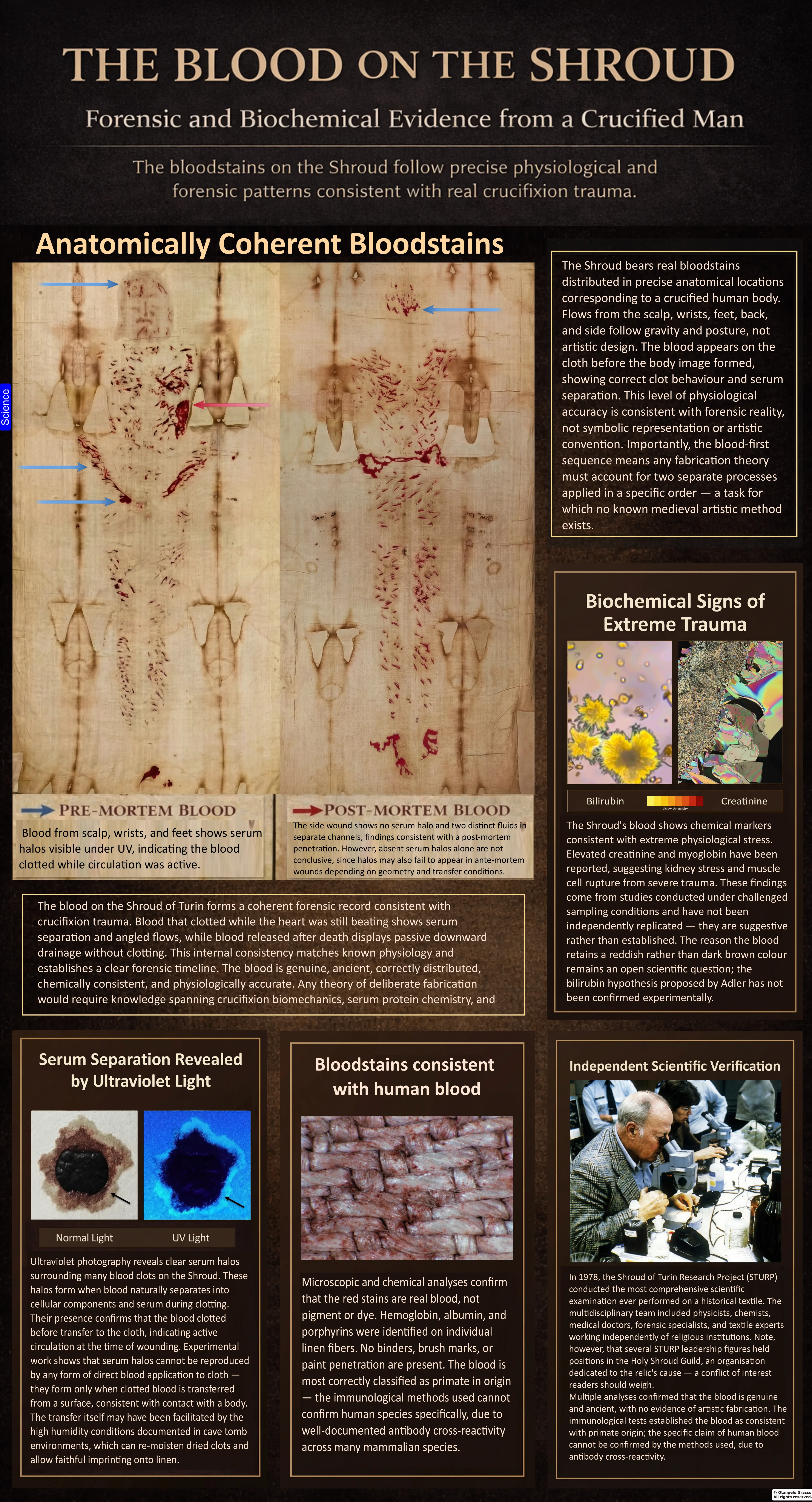
Reddish marks cross the linen at the head, wrists, feet, side and back. Investigators have reported blood-related chemical signatures in sampled stains, while other analysts have disputed how far those tests can go. The marks also invite a forensic question: do they record natural bleeding, deliberate application, or several processes at different times?
1. How the stains became a research question
For centuries, observers called the reddish-brown marks “blood” because of their appearance and their placement on a figure interpreted as wounded. Appearance alone cannot determine composition, however. The modern investigation asked whether chemical tests could distinguish blood residues from pigment, dye, or other material, and whether patterns on linen could provide evidence about how a fluid was deposited.
In 1973, a Turin commission supplied short threads from marked areas to a team at the University of Modena led by Giorgio Frache, with Eugenia Mari Rizzati and Emilio Mari. Their hematological tests, including benzidine and microspectroscopic procedures, were reported as negative. They cautioned that those results did not permit an absolute exclusion of blood in the material examined. The tested threads were tiny and centuries old, and the tests had practical limits; the finding belongs in the history but cannot by itself settle the question.1
In 1978, the Shroud of Turin Research Project (STURP) made a multidisciplinary examination using photography, ultraviolet imaging, spectroscopy, microscopy, X-ray fluorescence and adhesive-tape sampling. John Heller and Alan Adler later published chemical and spectroscopic tests on a tape sample from an alleged blood area. STURP’s 1981 summary stated that the stains were composed of hemoglobin and tested positive for serum albumin. These were important reported findings from particular samples and methods; they were not DNA identification of a named individual, nor a determination of when the marks were placed on the cloth.2, 3, 4
2. What can be seen on the cloth?
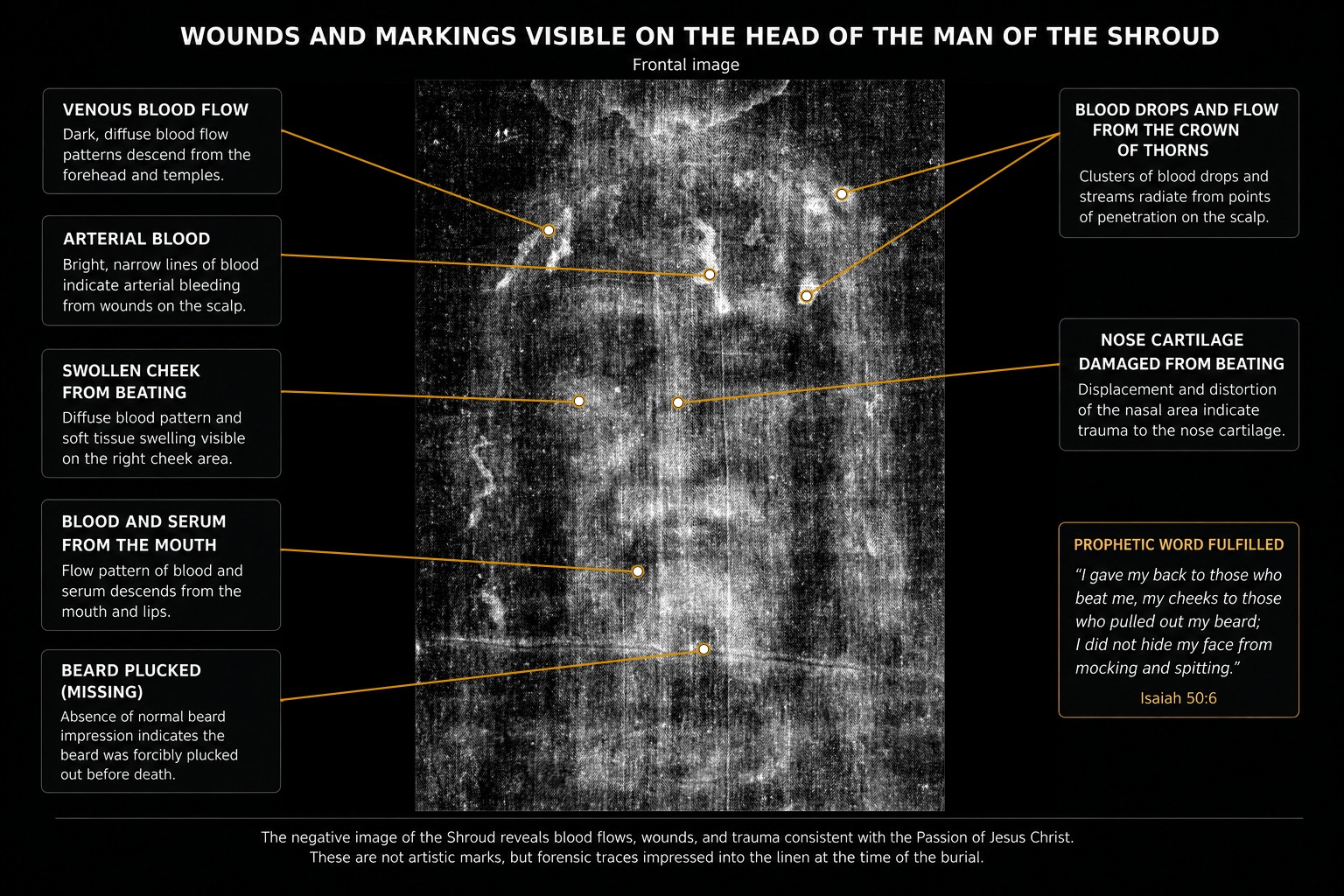
Stains appear on the scalp and forehead, wrists or hands, feet, side of the torso, and back and legs. Some are compact and irregular; others look like narrow rivulets. The image is faint and the linen has been damaged, repaired, handled and displayed. Contrast enhancement can make edges easier to inspect, but may also exaggerate color and texture.
It is useful to separate four levels of claim. First, a mark has a visible color, shape and location. Second, chemical or spectroscopic tests may identify substances in sampled material. Third, pattern analysis may assess how drops or flows could have formed. Fourth, an investigator may infer a wound or a historical event. The step from one level to the next requires argument and evidence. “Blood,” “arterial,” “venous,” “postmortem,” and “spear wound” are not labels written on the textile.
Common readings identify scalp marks as puncture wounds, paired back marks as scourging, wrist and foot stains as nail-related flows, and the large side stain as a piercing wound. Such readings are hypotheses to compare with medical, archaeological and physical evidence. The cloth contains no name for the depicted person, and stain morphology alone cannot establish a particular instrument, perpetrator or cause of death.
3. What are the stains made of?
Heller and Adler’s 1980 paper reported spectroscopic and chemical tests on a tape sample, including conversion of heme to porphyrin, and concluded that the material provided evidence for blood in the alleged blood areas. Their 1981 chemical investigation reported additional findings interpreted as blood proteins and bile pigments. STURP’s broader study considered optical spectra, fluorescence, elemental distribution and the interaction of colored material with linen fibres.2, 3, 5
These methods answer different questions. Spectral similarity to hemoglobin is not by itself a species test. A protein reaction is not unique to blood unless the assay is specific and cross-reactions are excluded. Iron detection alone does not establish hemoglobin, because iron-bearing particles can have other sources. The affirmative case rests on the reported convergence of several observations, not one decisive test. The original samples were minute, chemically heterogeneous and exposed to centuries of handling, fire, water and conservation; not every historical measurement can now be repeated on the cloth.
Walter McCrone’s microscopy and analytical work led him to argue that iron-oxide and vermilion pigment particles supported an artistic explanation. STURP reported that it found no pigments, paints or dyes on the fibrils sufficient to explain the body image, and Heller and Adler interpreted their own sampled reddish material as blood. The disagreement concerns the interpretation and significance of observed particles and tests. Pigment in some sampled material would not automatically explain every visible stain; blood-related compounds in selected samples would not prove every red mark consists exclusively of blood. The competing claims need to be represented clearly rather than reduced to a simple slogan.4, 6, 7
4. Species, blood group and DNA
Popular accounts frequently state that the Shroud’s blood is definitively human, type AB, male, or genetically linked to the Sudarium of Oviedo. Those conclusions exceed what is securely documented by modern forensic standards. Earlier serological reports assigned an AB group and human or primate origin. Kelly Kearse’s reassessments explain that aged stains, contamination, cross-reactivity and older polyclonal-antibody methods limit those identifications. Kearse concludes that the reported ABO designation is inconclusive and cautions that species testing can cross-react. The blood group should be described as a historical report, not a settled identification.8, 9
DNA is especially vulnerable to overinterpretation. A cloth displayed, handled, repaired, sampled and conserved over many centuries can accumulate biological material from people and environments. DNA detected on a fibre would not automatically belong to the person represented, or even to the original stain. A strong identification requires documented sample provenance, contamination controls, validated methods for degraded material, independent replication and evidence tying the genetic material to a stain rather than later contact. Claims of a particular donor’s sex, ancestry or relationship to another relic therefore remain unconfirmed.
5. What bloodstain-pattern analysis can do
Bloodstain-pattern analysis studies how drops, flows, transfers and impacts may form. In 2019, Matteo Borrini and Luigi Garlaschelli published experiments involving selected hand, forearm and torso marks. Using volunteers, simulated blood and a mannequin, they argued that some proposed flows did not fit the specific postures and scenarios tested. Their study challenged confident reconstructions that infer one exact body position from a still image.10
Those experiments did not recreate an execution, a wounded cadaver, removal from a cross, transport, contact with linen and burial as a single process. In 2024, Andreas Heger and colleagues tested movement-dependent blood patterns at the wrists using living volunteers. Some arm movements produced diverging flows resembling features attributed to the Shroud. The authors did not model a true nail tract, postmortem drainage, unnailing or transfer to the burial cloth. Both papers are useful because they make specific scenarios testable; neither is a complete reenactment of the Shroud’s proposed history.11
A stain rarely identifies one unique posture. Gravity, body position, rotation, contact with hair or skin, cloth pressure, drying and the order in which fluids arrive can redirect a flow. Pattern analysis can test defined alternatives under controlled conditions. A photograph alone cannot supply a complete event narrative.
6. Ultraviolet margins and blood-transfer experiments
Vernon Miller and Samuel Pellicori published ultraviolet-fluorescence photographs made during the 1978 examination. Several stains show pale fluorescent margins that are less apparent in ordinary photographs. Some researchers interpret these as serum associated with clot retraction: as a clot contracts, a lighter liquid fraction may separate from the darker red-cell-rich material and spread farther into absorbent cloth. The observed feature is a fluorescent margin; identifying it specifically as serum requires evidence beyond its appearance under ultraviolet light.12
Kelly Kearse tested ultraviolet illumination near 365 nanometres for visualizing serum patterns and conducted controlled blood-transfer experiments. His results suggest that clotting, drying and humidity can affect whether peripheral patterns form and transfer to cloth. This provides a testable physical mechanism for some halo-like features. It does not establish that every pale margin on the Shroud is serum, or that a mark was transferred directly from a corpse. Independent replication with controlled blood composition, characterized fabrics, blinded scoring and clear positive and negative controls would make the inference stronger.13, 14
Earlier experiments by Gilbert Lavoie and colleagues examined how the interval between clot formation and contact with cloth affected the transferred pattern. This work supports the general point that timing and material state matter. It does not uniquely determine when or how the Shroud stains formed.15
Miller and Pellicori’s original article includes ultraviolet photographs of the side-wound area and other stains. View the photographs and the authors’ captions. The source lets readers inspect the observation while keeping the “serum halo” explanation separate from the image itself.12
7. Can stains distinguish bleeding before and after death?
Forensic discussions distinguish bleeding while circulation continues from blood or fluid released after death. Clotting, serum separation, gravity, fibrinolysis and later disturbance can all affect a stain. Some Shroud interpretations assign certain flows to bleeding before death and others to passive postmortem drainage. The methodological question is whether the chemistry and geometry reliably discriminate those stages in an ancient textile.
The site author’s 2026 review proposes a composite scenario: clot formation and serum separation, followed by later fluid movement during handling, and transfer to linen while some material remained moist or was rehydrated. The author explicitly recognizes that single markers are not validated as definitive premortem or postmortem classifiers in ancient stains, that a serum-like margin does not date a stain, and that the full sequence has not been reproduced in one controlled reconstruction. It is a working, testable model, not a forensic determination of the time or cause of death.16
Claims that bilirubin or creatinine readings diagnose extreme trauma, asphyxia or renal failure should also be qualified. The author’s earlier biochemical synthesis presents such interpretations; the later critical transfer review says that several biomarker and microscopic assertions need independent confirmation. A claim in a synthesis paper is not the same as replicated clinical identification in authenticated, well-preserved blood from the Shroud. The author’s papers are downloadable below so readers can assess the arguments alongside the primary literature.17, 16
8. Blood, pigment and biological contact
McCrone argued that pigment particles supported an artistic explanation. Heller and Adler, STURP and other researchers argued that chemical and optical evidence in sampled reddish areas was consistent with blood. The alternatives need not be framed as though a historical cloth could contain only one material. The analytical question is whether detected materials explain the particular stains and their distribution.
A biological-contact model can account for blood-like marks and some fluid-transfer behavior, but does not by itself prove transfer directly from a crucified body. A maker could use genuine blood; blood and clear fluid could be applied sequentially; later contamination could add material; and the original marks could be changed by fire, water and handling. An artistic model must likewise account for reported blood chemistry, stain penetration and ultraviolet behavior, not merely point to pigment particles. A fair comparison states what each model predicts and tests those predictions against authenticated samples and controlled replicas.
9. What the blood evidence can establish
The Shroud’s reddish marks have been studied by chemical, spectroscopic, microscopic, ultraviolet and pattern-analysis methods. Heller and Adler reported blood-related chemical signatures; STURP summarized its findings as including hemoglobin and serum albumin; McCrone argued for pigment; and later experiments have investigated how posture, clot timing, ultraviolet fluorescence and humidity affect patterns. The result is a substantial but contested body of evidence.
It does not securely identify a named person, establish an AB blood group to modern standards, determine the donor’s sex or ancestry, prove a specific cause of death, or date the linen. Nor does a plausible transfer model authenticate the Shroud. The measured conclusion is narrower: sampled stains have yielded materials interpreted as blood-related, while the species, deposition history and relation between the stains and body image remain debated. Each claim should be weighed at the level its method supports.
References and research papers
- Giorgio Frache, Eugenia Mari Rizzati and Emilio Mari, “Relazione conclusiva sulle analisi di ordine ematologico effettuate sul materiale prelevato dalla Sindone” (1976), Turin commission research volume. The 1973 thread tests were reported as negative; the authors cautioned that the findings did not absolutely exclude blood. [Report transcription and context].
- John H. Heller and Alan D. Adler, “Blood on the Shroud of Turin,” Applied Optics 19, no. 16 (1980), pp. 2742–2744. [DOI and abstract] · [Reprinted paper].
- John H. Heller and Alan D. Adler, “A Chemical Investigation of the Shroud of Turin,” Canadian Society of Forensic Science Journal 14, no. 3 (1981), pp. 81–103. [Read the paper].
- John Heller, “A Summary of STURP’s Conclusions” (1981), the team’s press-conference summary. [Read the summary].
- E. J. Jumper, A. D. Adler, J. P. Jackson, S. F. Pellicori, J. H. Heller and J. R. Druzik, “A Comprehensive Examination of the Various Stains and Images on the Shroud of Turin,” in Archaeological Chemistry III, ACS Advances in Chemistry 205 (1984), pp. 447–476. [Read the paper].
- Walter C. McCrone and Christine Skirius, “Light Microscopical Study of the Turin ‘Shroud’ I,” The Microscope 28 (1980), pp. 105–113. [Publication record].
- Walter C. McCrone, “The Shroud of Turin: Blood or Artist’s Pigment?” Accounts of Chemical Research 23, no. 3 (1990), pp. 77–83. [DOI].
- Kelly P. Kearse, “Unanticipated Issues in Serological Analysis of Blood Species: The Shroud of Turin as a Case Example,” Forensic Science International: Reports 2 (2020), article 100073. [Open access paper].
- Kelly P. Kearse, “Inadequacies in Serological ABO Typing of Ancient Artifacts: The Shroud of Turin as a Case Example,” International Journal of Forensic Sciences 8, no. 3 (2023). [DOI and article].
- Matteo Borrini and Luigi Garlaschelli, “A BPA Approach to the Shroud of Turin,” Journal of Forensic Sciences 64, no. 1 (2019), pp. 137–143. [DOI] · [Author manuscript].
- Andreas Heger et al., “Further Experiments and Remarks Regarding the Possible Formation of Blood Stains on the Turin Shroud: Stains Attributed to the Nailing of the Hands,” International Journal of Legal Medicine 138 (2024), pp. 1573–1581. [DOI].
- Vernon D. Miller and Samuel F. Pellicori, “Ultraviolet Fluorescence Photography of the Shroud of Turin,” Journal of Biological Photography 49, no. 3 (1981), pp. 71–85. [Read the paper].
- Kelly P. Kearse, “Ultraviolet 365 as an Alternative Light Source for Detection of Blood Serum,” Journal of Forensic Sciences 65, no. 5 (2020), pp. 1716–1721. [DOI].
- Kelly P. Kearse, “The Effect of Humidity on Blood Serum Pattern Formation and Blood Transfer,” Journal of Forensic Science and Research 7 (2023), pp. 40–48. [DOI].
- Gilbert R. Lavoie, B. B. Lavoie, V. J. Donovan and J. S. Ballas, “Blood on the Shroud of Turin: Part II—The Importance of Time in the Transfer of Blood Clots to Cloth as Distinctive Clot Images,” Shroud Spectrum International 8 (1983), pp. 2–10. [Read the paper].
- Otangelo Grasso, “Blood Transfer, Serum Halos, and Postmortem Flow on the Shroud of Turin: A Critical Forensic and Hematological Narrative Review” (2026). Author’s testable model and its stated evidential limits. [Download the article].
- Otangelo Grasso, “A Forensic and Biochemical Analysis of the Bloodstains on the Shroud of Turin” (2026), author’s synthesis. [Download the article PDF] · “Pre-Mortem and Post-Mortem Blood on the Shroud of Turin” (2026), author’s synthesis. [Download the article PDF].
Further reading: Kelly P. Kearse, “Blood on the Shroud of Turin: An Immunological Review” (2012); Kearse, “Validation and Limitations in the DNA Analysis of Aged Bloodstains” (2023).