IN-DEPTH READING
The evidence in context
This chapter presents historical, scientific and medical interpretations of the Shroud. Some conclusions remain debated. A source note identifies the related work1.
The body is gone. Only its shadow remains.
The Task
No corpse lies on the steel table. No scalpel opens the body cavity. No tissue samples await the microscope. Instead, the pathologist confronts an image — a faint sepia stain on ancient cloth — and must work backward through time and biology to reconstruct what happened to a man in his final hours.
The wounds must speak where the voice cannot.
What makes this investigation forensically compelling is not merely that wounds are present, but that they configure themselves in ways medieval imagination would have been unlikely to conjure. Where centuries of sacred art showed nails through palms, the Shroud shows bloodstains consistent with wrist wounds. Where tradition depicted feet nailed separately, the cloth suggests a single spike through overlapped limbs. Where devotional images simplified suffering into stylized poses, this fabric captures the c haotic struggle of a man fighting for each breath. These are not the marks of artistic convention but of anatomical necessity — details that would remain unvalidated until the twentieth century brought cadaver experiments and forensic science to bear on th e mechanics of Roman execution.1
During the 1978 Shroud of Turin Research Project (STURP), forensic pathologists including Dr. Robert Bucklin conducted detailed analysis of the linen's bloodstain patterns and wound marks, working with high - resolution photography and direct examination of the cloth. This chapter synthesizes those findings and subsequent forensic analyses, beginning not with the cross but with what preceded it.
Before the Cross — The Protocol of Degradation
Before wood touched flesh, before iron pierced bone, the condemned man was stripped.
The Synoptic Gospels record this with characteristic brevity: they divided his garments among them. The passive voice obscures what was an active violation — the forcible removal of every thread of clothing, the exposure of the body to public view, the transformation of a person into an object.1 John's Gospel provides operational detail: a quaternion of four soldiers composed the typical execution squad, each entitled to a share of the condemned man's possessions as a grim perquisite of duty. The division was bureaucratic. Routine. Performed with the indifference of men who had done this before.
Image 12.1: A Passion scene showing Jesus being stripped of his garments by Roman soldiers before crucifixion. The deliberate exposure of the body was psychological warfare — using the honor -shame calculus of first - century Judean culture as an instrument of domination before any physical instrument was applied.
One garment disrupted the routine division — the tunic, seamless, woven in one piece, too valuable to tear. The soldiers cast lots for it. What appears as casual avarice becomes, within the scriptural framework, an unwitting enactment of Psalm 22:18 — they divide my garments among them, and for my clothing they cast lots — written a thousand years before the event, enacted without the soldiers knowing it.
This was not incidental cruelty but calculated procedure. In the symbolic universe of ancient Judaism, nakedness evoked primal shame, the reduction of person to mere flesh. Roman executioners understood this cultural calculus perfectly and weaponized it. T he man on the cross died many deaths. The stripping was the first. Everything that follows is clinical examination of what came after.
The Wrists — Where Anatomy Corrects Tradition
What the tradition said:
For over a thousand years, Western art spoke with one voice on this point: crucifixion nails pierced the palms. From Byzantine mosaics to Gothic manuscripts, from Renaissance paintings to Baroque sculptures, the iconography was consistent and universal. Ch rist's hands bore stigmata through the palms — visible, symmetrical, theologically charged with the symbolism of wounds that could be displayed, touched, verified. The tradition was so deeply embedded in devotional practice that it had acquired the force of revealed truth.
Image 12.2: A crucifixion scene showing Roman soldiers nailing Jesus to the cross. The artistic tradition consistently depicted nails through the palms — a convention that would not be challenged until Pierre Barbet's cadaver experiments in the 1930s raise d serious questions about its biomechanical plausibility.
What the Shroud shows:
The bloodstains originate not from the palms but from the wrists. Not a subtle difference. Not a matter of artistic interpretation. A fundamental anatomical correction to an error that had pervaded European sacred art for more than a millennium.
What anatomy confirms:
In the 1930s, Dr. Pierre Barbet brought cadavers into the conversation. His experiments were simple in conception and significant in implication. He drove nails through the palms of cadaver hands and suspended weight from them. The result was mechanical failure. The soft tissues of the palm — muscle, fascia, ligament — are biomechanically ill-suited to sustain the prolonged load of a suspended body without tearing. 2 Under tension, the flesh tears progressively until the nail pulls through completely. Barbet concluded that a man crucified through the palms alone could not be sustained in suspension. (Subsequent research by Frederick Zugibe suggested that certain bony structures of the hand may also provide some support, though this remains contested within the forensic literature.)
Barbet's experiments identified a region at the wrist as the structurally viable nail site: the space of Destot, a passage between carpal bones through which a nail could pass without fracturing them. He theorized that the nail would be held in place by th e surrounding bones and the strong ligaments of the wrist, providing a more structurally sound point of suspension than the palm.3 Unlike the palm, this location offered structural support capable of bearing the body's weight throughout the ordeal of crucifixion.
The forensic significance extends beyond biomechanical adequacy. A nail penetrating the carpal region of the wrist would be highly likely to traumatize the median nerve — one of the three major nerve trunks supplying the hand — depending on the precise ang le of entry. 2 The expected consequences include disruption of thenar muscle function, producing characteristic changes in thumb position. It should be noted that the precise neurophysiological mechanism remains a subject of debate: Barbet proposed that trauma to the me dian nerve would trigger thenar muscle contraction, drawing the thumb across the palm; however, authoritative anatomical references confirm that median nerve injury typically produces paralysis of the thenar muscles rather than spastic contraction, resulting in a flaccid thumb. 6 Zugibe further argued that the natural resting position of the hand in death offers a simpler explanation for the observed feature, demonstrating with anatomical models how the thumb falls naturally beneath the fingers when the wrist is nailed.4
Image 12.3: An illuminated folio from Jean Pucelle's Hours of Jeanne d'Évreux (c. 1324–1328, Paris), showing Christ nailed through the palms — consistent with medieval iconography but at odds with the forensic evidence reviewed in this chapter. The Shroud of Turin shows wrist wounds and the apparent absence of visible thumbs, details not found in known artworks of this period.
The Shroud's hand images show no visible thumbs. Both hands display four fingers clearly, but the thumbs are absent from view — not erased, not artistically omitted. Simply not visible. For centuries, this detail went unremarked or was attributed to artist ic choice. Barbet hypothesized that this absence was due to thumb retraction caused by median nerve trauma from the nail passing through the wrist — thumbs retracted into the palms, hidden beneath the fingers. Other forensic interpretations, including the natural resting position of the hand in death, have also been proposed to explain this feature.4
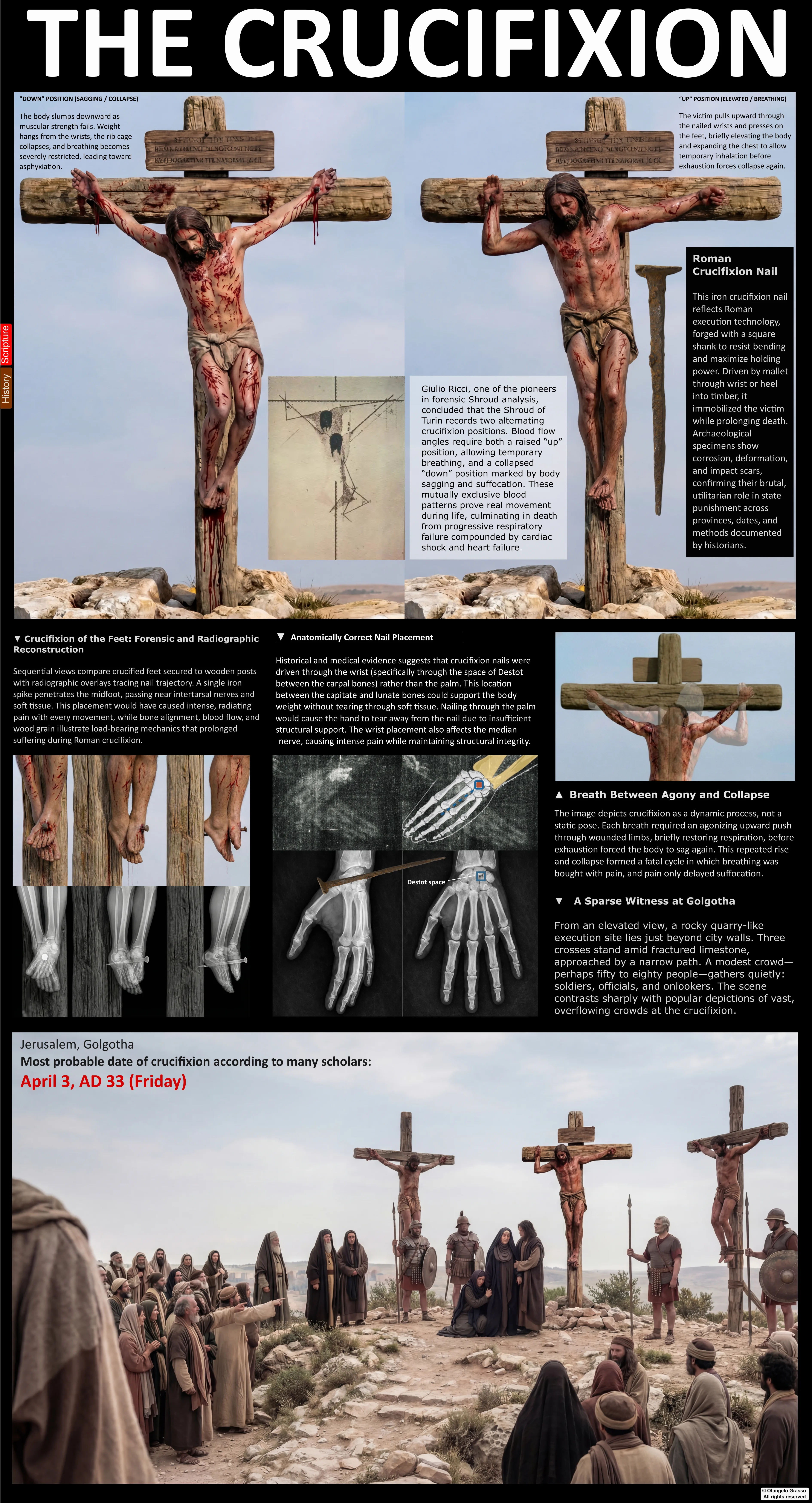
Image 12.4: The nail was driven through the wrist because the soft tissues of the palm cannot reliably support the weight of a suspended body, whereas the wrist — through the carpal region — provides a structurally stable anchoring point capable of bearing load. The Shroud reflects this by showing bloodstains originating at the wrist area rather than the palms, along with the apparent absence of visible thumbs whose precise cause remains debated in the forensic literature.
There is another dimension to possible nerve trauma that wound images cannot directly show: causalgia. Damage to major nerve trunks does not produce simple pain but a specific syndrome of burning agony that radiates through the entire limb. Unlike pain fro m tissue injury — which can be localized, managed, sometimes endured — causalgia is continuous, inescapable, and intensifies with any stimulation. Every movement on the
cross, every shift of weight, every breath-driven elevation would have sent cascades of searing sensation from the wrist wounds through the arms.
The bloodstains running down the forearms tell their own story. Rather than flowing in a single consistent direction, the blood tracks show multiple angles — some steep and nearly vertical, others more horizontal — creating a pattern of divergent flows fro m the wrist wounds. This pattern is consistent with forensic predictions from a victim alternating between two body positions. 4 When slumped, the victim's weight hangs from the wrist wounds and blood runs vertically. When the victim pushes upward on the nailed feet to draw a breath, the flow becomes more horizontal. The bloodstain geometry is consistent with dynamic changes in body position across the duration of crucifixion. It should be noted that Zugibe's volunteer experiments led him to question whether respiratory oscillation was the primary mechanism of death, proposing instead that hypovolemic and traumatic shock played a greater role — an interpretation that does not negate the positional blood -flow evidence but reframes its context.4
All of this — the wrist location, the absent thumbs, the angled blood flows — sits in a cloth lying in Turin. What the cloth preserves, the forensic examination reads.
The Feet — One Nail, Not Two
What the tradition said:
Medieval and Renaissance crucifixion scenes show feet nailed separately, positioned symmetrically, rendered with the visual clarity devotional art demands. Two wounds, two bleeding points, two distinct nails. The iconography was virtually universal. It ser ved the symbolic purposes of sacred art: clear, legible, emotionally accessible. What the Shroud shows:
The feet visible on the Shroud image are not positioned side by side. They appear partially crossed — overlapped. One foot placed atop the other.
What anatomy confirms:
Rigor mortis — the post-mortem stiffening of muscle tissue — locks the body in whatever position it occupied at or shortly after death. If the feet had remained fixed in strictly parallel alignment during crucifixion, rigor would have preserved that parall el positioning when the body was taken down. The feet would have remained side by side, just as they had been on the cross.
Image 12.5: The Shroud's foot image shows the feet in an overlapped position rather than side by side, consistent with one foot placed over the other and secured by a single nail. The bloodstain pattern originates from a unified dorsal wound with flow path s compatible with this stacked configuration, rather than two independent puncture sites that separate nailing would produce.
The overlapped position suggests a different configuration: a single nail penetrating both feet, with one foot placed atop the other. When the nail was removed and the body laid in the tomb, rigor mortis would have been consistent with maintaining some deg ree of this crossed arrangement.2 The foot position thus provides indirect evidence compatible with single-nail fixation — a detail that diverges from the two- nail depiction common in artistic tradition. It should be acknowledged, however, that Roman crucifixion practice was not rigidly standardized: the only confirmed Roman -era crucifixion victim known to archaeology, Jehohanan ben Hagkol (discovered 1968 in Jerusalem), shows evidence of nailing through the heel bone, demonstrating that the specific anatomical details preserved on the Shroud cannot be treated as representing a single invariable protocol.
Establishing that one nail likely secured both feet raises a more technically challenging question: what anatomical path did that nail follow? Barbet proposed an intermetatarsal route — the nail passing through the space between the long metatarsal bones o f the forefoot. This path had anatomical elegance, but biomechanical analysis suggests complications: an intermetatarsal path alone might lack sufficient structural stability to support the body's movements during crucifixion.
More recent forensic and anatomical research has hypothesized that the nail may have followed a more complex trajectory through the overlapped feet. When one foot is placed atop the other, the nail could penetrate through different bone regions of each foo t — potentially through the tarsal region of one foot, providing stable anchoring, then continuing through the metatarsal space of the other. Such models attempt to reconcile the biomechanical requirement for stability with the anatomical constraints on nail placement.
A substantial bloodstain appears near the right heel on the Shroud, prompting some researchers to argue for a heel spike in addition to the dorsal foot wound. But the stain's diffuse character and irregular borders can be interpreted as consistent with pos t-mortem drainage — blood pooling in the foot cavity after death, then draining toward the heel when the body was laid horizontal and wrapped in cloth.1 Capillary action in the linen amplified this drainage, spreading it into a stain larger than the wound that produced it. This interpretation offers a parsimonious explanation and requires no additional nailing, though scholarly debate on this point continues.
The foot wounds parallel the wrist wounds in a crucial respect: they represent forensic detail that diverges from artistic convention while aligning with biomechanical necessity. These are not features born of religious imagination. They are read from phys ical evidence — the kind of details that emerge when cloth contacts an actual crucified body rather than when an artist paints an idealized one.
The Scourge Marks — The Preliminaries of Death
What the tradition said:
Devotional art of the crucifixion includes the scourging as a narrative episode — theologically significant, visually powerful — but typically renders the wounds with the restraint that sacred art demands. Stylized stripes. Symbolic suffering. The visual v ocabulary of martyrdom rather than the clinical record of a specific weapon.
What the Shroud shows:
The dorsal image records approximately 100 –120 individual wound marks. Each displays a characteristic dumbbell shape — two circular or elliptical contusions separated by a bridge of abraded or lacerated tissue.
They cover the back, buttocks, and posterior legs — essentially the entire rear surface of the body from shoulders to calves, applied in a pattern suggesting one or two executioners working systematically, moving the whip across the victim's body in overla pping strikes to ensure complete coverage.
What anatomy and archaeology confirm:
The morphology of these marks is widely considered characteristic of the impact pattern produced by a Roman multi -thonged whip with leather straps tipped with small paired metal weights, an instrument type attested archaeologically and commonly referred to in modern scholarship as the flagrum taxillatum .5 It should be noted that the term flagrum taxillatum itself does not appear in Roman sources; it is a sixteenth - century scholarly coinage by Justus Lipsius, subsequently adopted in Shroud research literature. The weapon type, however — a multi-thonged whip with metal attachments producing paired impact poin ts — is archaeologically confirmed. When such a weapon strikes the flesh, each pair of metal weights creates two impact points, with the connecting thong tearing the tissue between them. The result is the dumbbell lesion visible throughout the Shroud's dorsal image.
The sheer number — well over a hundred distinct lesions — indicates a flogging that went far beyond token punishment. This was scourging that approached lethal intensity, that transformed the back into a topography of trauma.
A flogging of this severity can initiate a cascade of physiological crises. Each strike breaks skin, lacerates muscle, ruptures capillaries. Multiply that trauma by a hundred, and the cumulative effect can be physiologically severe: massive blood loss leading to hypovolemic shock; acute systemic inflammation; severe pain generating neurogenic shock; and profound physical weakness.5 The victim who survives scourging and is taken to the cross is already in extremis — blood pressure dropping, heart rate elevated, strength depleted, body beginning to shut down. The pre - weakening was not accidental but integral to Roman execution protoco l: the scourge marks on the Shroud represent the opening phase of a coordinated procedure, one that would significantly affect the victim's physiological state during every minute on the cross.
And each of those approximately 100 –120 dumbbell-shaped wounds — with their serum halos, their precise morphology, their systematic distribution — is preserved in a cloth. The pathologist who examines it is examining evidence that has been sealed in linen for centuries. The clinical pattern and the ancient cloth are inseparable.
Synthesis — The Architecture of Authenticity
Step back from the individual wounds and consider the forensic evidence as an integrated system.
The scourge marks record pre -crucifixion flogging administered with a specific weapon type, distributed in a pattern consistent with systematic application. The wrist wounds show nail placement through a mechanically favorable anatomical location in the ca rpal region, with the consequent absence of visible thumbs — a detail whose precise mechanism remains debated but whose departure from artistic convention is not in question. The foot wounds are consistent with a single -nail configuration supported by rigo r mortis positioning. The bloodstain patterns are consistent with body position changes during crucifixion — the dynamic struggle of the victim recorded in the geometry of fluid mechanics.
These elements do not simply coexist. They form a coherent forensic narrative.
To attribute this image to medieval fabrication would require proposing that a fourteenth -century artisan either possessed, or had access to, practical or artistic knowledge not common in mainstream devotional art of the time. The forger would need to understand that nails must penetrate the wrist rather than the palm — a fact that contradicted the overwhelming majority of contemporary artistic tradition and that would not be subjected to systematic experimental examination until Barbet's work in the 1930s. (It should be acknowledged that a practically minded forger might conceivably have arrived at wrist nailing through direct observation or relic study, rather than anatomical theory, and this possibility cannot be entirely excluded.)
They would need to produce a detail — the non-visibility of the thumbs — that was inconsistent with prevailing iconography and lacked any devotional motivation, and whose precise forensic explanation remains debated even today. They would need knowledge of blood flow patterns that vary with body position during crucifixion — biomechanics not systematically developed until the twentieth century. They would need familiarity with the specific dumbbell wound morphology produced by a Roman multi-thonged whip with paired metal weights — a pattern whose diagnostic significance was not recognized until modern forensic analysis.
Each element individually might be explained. Together they form a coherent pattern that warrants explanation.
One significant counterpoint must be engaged directly: in 1988, three independent radiocarbon dating laboratories (at Arizona, Oxford, and Zurich) analyzed samples from the Shroud linen and produced a calibrated date range of approximately 1260 –1390 CE, placing the cloth's origin in the medieval period with 95% confidence.7 This finding remains the most significant single scientific test performed on the artifact. Whether one accepts these results as definitive — or believes them to be complicated by contamination, sampling from a later repair, or other factors — any authenticity-focused analysis must engage with them. The forensic wound
evidence reviewed in this chapter cannot, by itself, resolve the dating question; it addresses the character of the wounds recorded, not the age of the linen on which they appear.
Image 12.6: A crucifixion scene showing three men on crosses atop a barren hill, observed by a modest crowd and Roman soldiers. Against this historical backdrop the Shroud's integrated wound system — wrist nailing, single-nail foot fixation, systematic sco urging — presents a forensic account that diverges markedly from the available artistic tradition.
Against this backdrop, the contact hypothesis provides a parsimonious explanatory framework. If the linen wrapped an actual crucified body, all the forensic details follow naturally. The wrist wounds are in the carpal region because that is where Roman exe cutioners drove nails to support the body's weight. The thumbs are absent because of processes attendant on wrist nailing, whatever the precise mechanism. The blood flow patterns show multiple angles because the victim's position changed during crucifixion. The scourge marks show the characteristic dumbbell pattern because they were made by a multi -thonged whip with paired metal weights. The contact hypothesis explains not only the presence of wounds but their precise characteristics — the absent thumbs that no medieval artisan depicted, the blood flows that are consistent with the victim's struggle for breath, the systematic scourge pattern that matches a specific Roman weapon type whose diagnostic morphology was not recognized until the modern era.
Image 3.2. This forensic reconstruction depicts the crucified body with detailed blood flow patterns traced across the arms, torso, and lower body, showing the gravitational and anatomical pathways of blood seepage from crucifixion wounds. The overlaid gri d and anatomical markings document the precise distribution and direction of bloodstains on the Shroud, providing forensic evidence of the body's position and the physiological consequences of crucifixion trauma.
Image 3.3 & 3.4: The depiction showing the two positions of crucifixion — slumped downward and raised upward — illustrating the up-and-down movement associated with the breathing process. The divergent blood flow angles on the Shroud's wrist stains are consistent with this os cillation: positional changes during survival encoded in fluid dynamics and preserved in linen.
The wounds are witnesses. They tell a story of systematic brutality, of anatomical precision in execution, of a man who suffered according to a
specific historical protocol of Roman military violence. They were inflicted, endured, and recorded. They are the testimony of trauma, preserved in blood and linen, waiting for forensic science to develop the tools to read what they have been saying all along.
The wounds tell us how this man was killed. The next question is how long it took — and why it took far less time than Rome expected.
References
1. Bucklin, R. (1982). The Shroud of Turin: Viewpoint of a forensic pathologist. Shroud Spectrum International , December, 3 –11. Link. (Forensic pathological analysis of the Shroud image by the STURP team's forensic pathologist, detailing observations on wound placement, blood flows, and the mechanism of crucifixion death — the primary source for the overall wound taxonomy and the 1978 STURP examination framework applied in this chapter.)
2. Barbet, P. (1953). A Doctor at Calvary: The Passion of Our Lord Jesus Christ as Described by a Surgeon . P. J. Kenedy & Sons. Link. (The pioneering medical study of crucifixion based on Shroud evidence; contains Barbet's cadaver experiments establishing the biomechanical inadequacy of palmar nailing, identifying the carpal region of the wrist as the structurally viable nail site, and proposing the single-nail foot fixation model based on overlapped foot position and rigor mortis evidence.)
3. Wikipedia contributors. (2024). Space of Destot. In Wikipedia, The Free Encyclopedia . Link. (Reference article describing the anatomical space in the wrist relevant to the chapter's discussion of wrist nailing. For primary anatomical description, see also Barbet (1953), pp. 101–115, and Standring (2021), Section 6.)
4. Zugibe, F. T. (2005). The Crucifixion of Jesus: A Forensic Inquiry. M. Evans and Company. Link. (Modern forensic reassessment of crucifixion providing detailed analysis of blood flow patterns and wound mechanics on the Shroud; primary authority for the divergent -angle blood flow interpretation as a record of the victim's positional oscillation. Als o contains Zugibe's challenge to Barbet's median nerve / thenar spasm mechanism, his demonstration of the natural resting position of the hand, and his volunteer experiments questioning the asphyxiation model of crucifixion death.)
5. Edwards, W. D., Gabel, W. J., & Hosmer, F. E. (1986). On the physical death of Jesus Christ. Journal of the American Medical Association, 255(11), 1455–1463. Link. (Landmark medical analysis of the crucifixion process, detailing the pathophysiology of scourging and crucifixion; primary source for the wound morphology consistent with a Roman multi- thonged whip with paired metal weights, the physiological cascade fro m scourging to hypovolemic shock, and the biomechanical framework applied to the Shroud's dorsal scourge mark distribution.)
6. Standring, S. (Ed.). (2021). Gray's anatomy: The anatomical basis of clinical practice (42nd ed.). Elsevier. (Definitive anatomical reference describing the median nerve's distribution to the thenar muscles and confirming that injury produces motor paralysis rather than spastic contraction. See also Zugibe (2005), for application to crucifixion forensics.) Link.
7. Damon, P. E., et al. (1989). Radiocarbon dating of the Shroud of Turin. Nature, 337, 611–615. Link. (Landmark study reporting the results of independent radiocarbon dating by laboratories at the University of Arizona, the University of Oxford, and ETH Zurich, producing a calibrated date range of approximately 1260 –1390 CE for the Shroud's linen at 95% confidence.)
THE WIDER DEBATE
Wounds of the wrists, feet and side
The first thread argues that nails passed through the wrists (the space of Destot), that the feet were fixed with one nail through each, that the side wound is about 5 cm wide and matches a Roman lancea, and that the legs were not broken, in line with John 19:33–36.
Source: the two catsboard threads of the Otangelo Grasso forum, part 1 and part 2. These are the author’s arguments, presented here as such, not established findings.
Explore the author’s additional material: The Shroud of Turin, part 1 · Part 2. The book’s bibliographic references are included where available.