IN-DEPTH READING
The evidence in context
This chapter presents historical, scientific and medical interpretations of the Shroud. Some conclusions remain debated. A source note identifies the related work1.
Ray Rogers did not believe in the Shroud of Turin.
He was a chemist and thermal analyst from Los Alamos National Laboratory — a man who had spent his career applying thermal analysis
techniques to explosives and energetic materials research, where the universe did not care what you believed and molecules did not lie. When he joined the 1978 Shroud of Turin Research Project — a team of approximately thirty-three scientists and support p ersonnel who brought seven tons of equipment to Turin — he came with the quiet confidence of a man who expected to be home by Christmas with a clean answer. The cloth was probably a medieval painting, or a clever contact print, or some combination of the two. He would show his colleagues how it was done, pack up his spectrometer, and catch his flight.
He returned to active investigation of the Shroud in his final years, ultimately engaging with the problem across portions of the period 1978– 2005.
The Shroud of Turin is that kind of object. It does not reward casual dismissal, and it does not reward credulous acceptance. It sits at the precise, uncomfortable intersection of physics and theology, of forensic chemistry and medieval history, and it has been doing so for at least seven centuries — deflecting every attempt to reduce it to something simple, something explainable, something that would let a thoughtful person stop thinking about it.
This chapter is about the physics. Specifically, it is about a question that emerges from the data with the force of a logical theorem: whatever made the image on this cloth, it was not any known process that has yet been shown to satisfy all of its measur able constraints simultaneously. That conclusion is not a leap of faith. It is the residue left behind after major conventional mechanisms have been systematically tested and found wanting against the evidence. It is what remains when the well-studied has been set aside, and the inexplicable has not yet been explained.
To understand why, we need to start at the most basic level of all: a single thread.
I. The Molecular Transformation: What Color Is Made Of
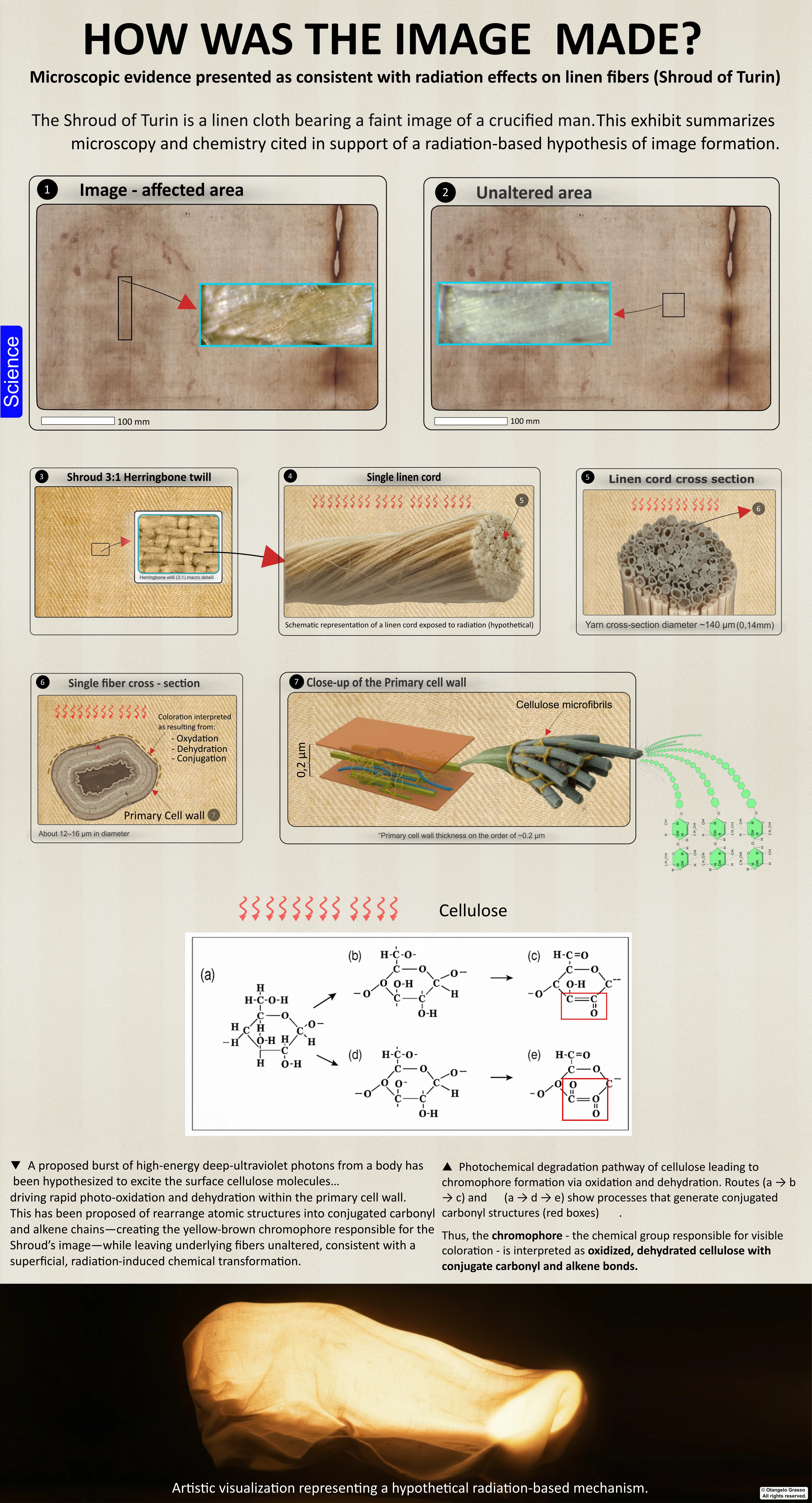
Pull a single fiber from the image area of the Shroud — if you were permitted to, which you are not — and place it under a scanning electron microscope. What you would see is not paint. It is not dye. It is not any foreign substance at all. The fiber itself has changed color. More precisely, the outermost layer of the fiber has undergone a chemical transformation, aging decades or centuries ahead of the surrounding material, as if someone had pointed a very precise clock at individual threads and fast - forwarded it.
Chemical analyses, from the 1978 STURP investigation to subsequent nano-scale studies, converge on a three-stage molecular reaction sequence that reads less like an artistic process and more like a chemistry textbook entry on accelerated degradation.1,2
Stage 1: Dehydration. Water molecules are stripped from the cellulose polymer chains that give linen its structure. This is not a gentle process. Photochemical dehydration of cellulose via direct bond cleavage requires deep-ultraviolet photons — wavelengths between 200 and 280 nanometers — carrying energies of approximately 4.8 electron volts per bond. For context, the photons in ordinary visible light carry about 2 electron volts. Whatever did this was operating in an energy register that sunlight barely touches.
Stage 2: Oxidation. With the hydroxyl group stripped away, the exposed carbon atom reaches out to oxygen in the surrounding air, forming a carbonyl group — the molecular signature of an aldehyde or ketone. This is the chemistry of yellowing paper, of aging fabric, of a document left too long in a sunny archive.
Stage 3: Conjugation. The final transformation is the most visually consequential. Normal single carbon -carbon bonds convert into an alternating network of single and double bonds. This is called conjugation, and its effect is optical: the resulting molecular structure — a chromophore — absorbs blue light at wavelengths around 400 to 430 nanometers, and so the fiber appears straw-yellow to brown to the human eye. The Shroud's image is not a color applied to the cloth. It is the cloth itself, selectively aged.
This last point deserves to sit with you for a moment. Aging, as any conservator will tell you, is indiscriminate. A document left in the sun turns yellow uniformly — every page, every margin, every blank space. What has happened to the Shroud's image fibe rs is aging that is not indiscriminate at all. It is aging with the precision of a surgeon, browning certain fibers in a pattern that encodes the three -dimensional shape of a human body, while leaving fibers a fraction of a millimeter away in their original, pristine state. As if a book left in the sun browned only the letters of a specific poem, while every surrounding page remained white.
The Energy Budget: A Small Number with an Impossible Delivery
Here is where the story becomes quantitative, and the numbers are worth following carefully, because they contain the heart of the puzzle.
The Shroud measures 4.4 meters by 1.1 meters. The body image covers roughly 1.21 square meters of that surface. But the transformation goes only 0.2 micrometers deep — a fifth of a millionth of a meter, less than a hundredth of a human hair. The affected v olume works out to approximately 0.024 cubic centimeters — a material weighing about 0.036 grams.
Within that tiny, tiny mass lie approximately 1.4 × 10²⁰ cellulose molecules and approximately 8.4 × 10²⁰ carbon atoms. Conservative
estimates suggest perhaps one in every thousand of those carbon atoms underwent the critical transformation from single to double bond — each requiring approximately 3.5 electron volts of energy.
These values are model-dependent estimates rather than directly measured quantities, but they establish the correct order of magnitude for assessing the energy delivery challenge.
The challenge is not, it turns out, the total quantity of energy. A sequential raster-scan of the full image — the author's own preferred model, developed in Section V — requires approximately 29 to 36 kilojoules in total delivered energy. 4 This is deeply unimpressive on its face. The problem — the one that has consumed physicists and chemists for decades — is how that energy must have been delivered. And for that, we need to understand what the image actually is, in physical terms, before w e can understand why creating it should be so difficult.
II. The Combinatorial Lock: Twenty-Five Keys Required
Imagine a safe that does not have one combination — it has twenty-five, all of which must be entered simultaneously. This is the situation facing any scientist attempting to explain the Shroud's image. Over decades of multidisciplinary analysis, the eviden ce has crystallized into a set of physical and chemical requirements — a combinatorial lock that any valid theory must pick in its entirety, not selectively. Fail even one, and the theory is eliminated.
Recent work of mine (2025) synthesized this scattered literature into a systematic framework of twenty -five empirically testable requirements, organized into four tiers of evidential weight. 4 They are not articles of faith. They are measurements.
Tier 1: Fundamental Physical Properties (11 Requirements)
R1 — Extreme Superficiality. The image coloration penetrates only the outermost 0.2 to 0.6 micrometers of a fibril's primary cell wall — less than one percent of the fibril's total diameter. The interior of each fiber, its lumen, and every fiber lying beneath t he surface layer are completely pristine.5
Here lies the first paradox, and it is a striking one. Deep ultraviolet radiation typically penetrates to depths orders of magnitude greater than the 0.2 -micron image layer under laboratory conditions. The Beer - Lambert law, one of the foundational equation s of spectroscopy, tells us that photons attenuate gradually through a medium, following an exponential decay. They do not stop at a predetermined depth like a polite guest who won't cross a threshold. They penetrate until they are absorbed or scatter away. Yet on the Shroud, the image ends at 200 nanometers as if the process understood exactly when to stop.
R2 — Single-Sided Imaging. No coherent image exists on the reverse side of the cloth. Some isolated fiber-level effects have been identified, but no duplicate image — which any penetrating radiation or diffusing chemical would inevitably produce.2
R3 — Three-Dimensional Encoding. This is perhaps the most extraordinary property of all. The image's density — how dark or light a given area appears — varies in precise, quantitative correlation with the distance between cloth and body. Areas where the cloth w as close to the surface (the nose, the cheekbones, the knuckles) are darkest. Areas where the cloth was farther away (the eye sockets, the sides of the torso) are progressively lighter. It is not artistic chiaroscuro — the painterly technique of implying depth through shading. It is a genuine spatial map, encoded in chemistry.3
The VP -8 Image Analyzer, was used in various imaging applications including NASA -related projects, was applied to photographs of the Shroud in the 1970s. When ordinary photographs — portraits, paintings, any flat image — are fed into the VP -8, they produce distorted, meaningless blobs, because flat images contain no genuine distance data. The Shroud produced a dimensionally accurate, anatomically coherent three-dimensional model of a human body. The image had been encoded with spatial information that no known artistic process could have placed there.
R4 — Photographic Negative Properties. The image on the cloth is a negative: light areas in the cloth correspond to the body's raised surfaces, and dark areas to its recesses — the inverse of what you would expect from a contact stain. When an Italian lawyer an d amateur photographer named Secondo Pia processed the first photographic plates taken of the Shroud in 1898, he almost dropped them in his darkroom. His photographic negative revealed a positive — a luminous, detailed portrait of a face staring back at him from the glass. He assumed he had made an error. He had not. The Shroud is itself a negative, and only in 1898 did humanity possess the technology to recognize that fact.2
R5 — Intrinsic Oxidative Cellulose Chemistry. FTIR spectroscopy, Raman analysis, and X -ray photoelectron spectroscopy all confirm the same thing: the image is consistent with carbohydrate oxidation and dehydration, and nothing else. No pigments. No binding medi a. No dyes. No varnish. No protein-based paint. Nothing foreign is present in the body image above detection limits. 1 STURP's chemical analyses, summarized by Schwalbe and Rogers (1982), found the image consistent with a cellulose oxidation and dehydration phenomenon — with the overall 1981 STURP summary concluding the image "remains a mystery" as to its formation mechanism.
R6 — Binary Threshold Behavior at Fibril Scale. Under the microscope, the image dissolves into something astonishing. Individual fibrils — the finest structural unit of the linen thread — are either fully colored or fully uncolored. There are no gradients within a single fibril, no partial staining, no subtlety at the microscopic level. The continuous tones of the image, the impression of gradual light and shadow that makes it look like a picture, are a statistical illusion: they result from varying densities of fully- switched fibrils, exactly like the halftone dots of a printed photograph that resolve into continuous tones at reading distance.6
Dr. John Jackson estimated approximately 5.93 million such binary information elements encoded in the cloth. This is not image formation in any conventional sense. It is image encoding — the translation of three - dimensional spatial data into a precise, durable chemical representation at the limit of molecular resolution.
R7 — Stochastic Fibril-Level Mottling. At the finest scale, there is visible randomness in which fibrils were colored — a "shot noise" effect, as if the coloring process operated statistically near a threshold rather than deterministically.6
R8 — Selective Surface Fibril Targeting. Within any given yarn bundle, only the exposed surface fibrils are affected. Fibrils buried in the interior of the thread, shielded by their neighbors, remain completely uncolored.2
R9 — Absence of Thermal Damage. The image shows no pyrolysis — no partial burning — and no carbonization of any kind. When examined under ultraviolet light, scorch marks fluoresce visibly. The body image does not. The chemistry of the image is oxidation and conjugation, not the thermal degradation pathway that heat would produce.7
R10 — Spatial Resolution (~5 mm). Anatomical detail resolves consistently at approximately five millimeters across the cloth surface — precise enough to distinguish individual fingers, individual wound marks, the contours of a face.3
R11 — Frontal/Dorsal Maximum Density Parity. The front and back images of the body exhibit comparable maximum optical densities. Whatever process operated on the front of the cloth also operated equivalently on the back — suggesting a single, uniform formation mechanism rather than, say, gravity-dependent staining.8 Tier 2: Blood-Image Integration (6 Requirements) Before moving forward, it is worth pausing on the blood — because the relationship between the blood and the body image is itself one of the most forensically significant features of the cloth, and it imposes its own set of constraints on any formation theory.
R12 — Blood Temporal Precedence. The blood and serum films overlie the image fibers. Ultraviolet fluorescence imaging, which makes the
serum halos around each bloodstain visible as a kind of aureole, strongly indicates that the blood was deposited before image formation. The image does not continue beneath the bloodstains. It stops at their edges.7,10
R13 — Blood-Image Non -Interference. Whatever process formed the image left the clot rims and serum borders perfectly intact. The image ends cleanly at blood boundaries, as if the image -forming energy was blocked by the blood — or knew to stop.10
R14 — Human Blood Biochemical Signatures. The blood chemistry has been extensively analyzed. Immunochemical testing confirms the presence of heme, albumin, and immunoglobulin G — markers consistent with human blood, not animal blood or pigment.11
R15 — Anatomical Blood -Image Correlation. The blood flows and wound marks align anatomically with the underlying body image. They are not random splatters — they are positioned where wounds on the imaged body would have produced them, with directionality consist ent with the orientation of the figure.10
R16 — Off-Image Bloodstain Presence. Blood evidence — scourge marks and other contact transfers — appears outside the body image zone entirely, in areas where there is no body image at all. This confirms that blood was deposited by a physical body lying in conta ct with the cloth, independently and prior to any image-forming event.7
R17 — Absence of Putrefaction Evidence. No biological markers of advanced decomposition appear anywhere on the cloth. No putrefaction staining, no chemical signatures of tissue breakdown, no artifacts of bacterial activity. If a body produced this image, it was wrapped for fewer than approximately forty hours — before the chemistry of decay had time to begin its own, very different kind of transformation.8
Tier 3: Geometric, Anatomical & Wrapping Precision (7 Requirements)
R18 — Predominantly Orthogonal Projection. The image shows minimal perspective distortion. It is consistent with radiation or emission traveling perpendicular to the cloth surface — in parallel, collimated rays — rather than the diverging, angle-dependent projection you would get from a body pressing against fabric.2
R19 — Accurate Human Proportions. The body's anthropometry is consistent with human norms. It is not stylized, not idealized, not elongated in the manner of medieval religious art. It is simply a man, with the proportions of a man.3
R20 — Wrapping-Consistent Image Distortions. Here is one of the most counterintuitive requirements. The Shroud image exhibits certain distortions in the hands, calves, and torso that are consistent with a cloth
draped over a three -dimensional form, while simultaneously lacking the gross wraparound distortions expected from a simple physical contact imprint. This complex geometry — both compatible with a draped cloth yet inconsistent with full contact imaging — is a key feature any formation hypothesis must address.12
R21 — Absence of Lateral (Side) Images. If the cloth was wrapped over a body and the image -forming process was a diffusing gas, or a contact stain, or anything that spreads or wraps, you would expect to see images on the sides of the cloth — where the fabric cur ved around the flanks. There are none.3
R22 — Natural Edge Transition. The image fades gradually at its boundaries. There is no sharp edge, no stencil cutoff, no hard border as you would expect from a template or a contact print.3
R23 — No Diffusion Signatures. There are no halos, no capillary spread patterns, no blurring at boundaries — none of the marks that any liquid or vapor process leaves behind as it migrates through fabric.2
R24 — Forensic Corpse Alignment. The body's posture is consistent with rigor mortis. The fingers are in fixed flexion. The foot positioning matches a crucifixion victim removed from a cross. This is not an artist's idealized figure in repose — it is a forensic r ecord of a body in the specific configuration that death and rigor would have fixed it.1,12,2
Tier 4: Uniformity & Cloth-Topology Independence (1 Requirement)
R25 — Fold-Independent Imaging. Image intensity is determined solely by cloth-to-body distance. Folds, creases, wrinkles in the fabric — local topographic features of the linen itself — have no effect on image density whatsoever. The image -forming process appear s to have been entirely indifferent to the cloth's own surface structure, responding only to the geometry of the body beneath it.3
Twenty-five requirements. Twenty -five locked doors. Any theory that cannot open all of them simultaneously is not a partial solution — it is no solution at all.
Before these twenty -five requirements are applied as an elimination framework in the section that follows, one structural question about the framework itself must be named — because its answer determines how much weight the elimination can legitimately carry.
The requirements are presented here as the crystallised residue of decades of multidisciplinary analysis: measurements, not beliefs, built up empirically as investigators documented what the image actually is. That characterisation may be entirely accurate . But the framework was synthesised and codified in its current form in a single 2025 publication
by this book's author — working, necessarily, in full knowledge of the image properties the requirements describe and of the candidate theories that were to be tested against them. This creates a methodological question that the framework's use as a truth -preserving filter requires answering: were the twenty -five requirements established independently of the theories they are used to eliminate, or were some of them formulated — at least in part — in response to those theories?
The distinction matters because a set of criteria built up incrementally, in response to each new hypothesis, can become unfalsifiable by design. If a new naturalistic proposal explains properties R1 through R24, a twenty - fifth requirement can always be ad ded that the proposal fails. The framework then eliminates every hypothesis not by the force of the evidence, but by the elasticity of its own boundaries. This is not a charge being levelled here — it is a structural risk that any post-hoc consolidation of scattered criteria carries, and it is the risk that the framework's critics will identify.15
The appropriate response is not to abandon the framework — the individual requirements are empirically grounded and most have extensive independent documentation in the literature cited throughout this chapter. It is to be explicit about which requirements were established a priori — documented before any specific formation hypothesis was proposed and tested — and which emerged post-hoc in response to the shortcomings of particular theories. Several of the Tier 1 and Tier 2 requirements (R1, R3, R5, R12) have robust, independently published documentation predating the 2025 synthesis. Others, particularly in Tier 3, rely more heavily on the synthesis itself. Making this distinction transparent would strengthen rather than weaken the framework's use, because it would allow the reader to assess which eliminations rest on the most secure independent ground and which depend more directly on th e synthesiser's own analytical choices. The combinatorial lock is genuinely formidable. Its full force is best conveyed when its provenance is fully visible.
III. The Usual Suspects: Everyone Gets a Turn
Science proceeds by elimination as much as by discovery. Over a century of investigation has produced a long list of proposed mechanisms for the Shroud's image — and a longer list of reasons why each of them fails. Think of what follows as a lineup, where each suspect gets a fair hearing before the evidence exonerates or convicts them. The Painter. Start with the simplest hypothesis: a medieval artist with a brush and pigments created a remarkable religious artifact. It is the explanation that requires the least extraordinary assumption. It fails immediately and comprehensively. Pigment does not sit in a 0.2 -micron surface layer — it penetrates, it pools between fibers, it leaves binding media (R1, R5). Brushwork creates directional patterns in the fibers. Most fatally, no artist in the fourteenth century — or any century before 1898 — could have painted a photographic negative that resolves into a
coherent positive only when photographed. The photographic negative property (R4) alone eliminates the painter, because it requires a concept that did not exist.
STURP's chemical analyses found no pigments, paints, dyes or stains in the body -image fibers, and concluded the image is consistent with a cellulose oxidation phenomenon. 2 The painter is acquitted. Or rather, disqualified entirely — not because of insufficient motive, but because the cloth simply does not contain paint.
It is worth acknowledging here the work of Walter McCrone, the STURP microscopist who dissented from the team's majority conclusion. McCrone published findings in the Microscope journal (1980 –1981) identifying iron-oxide particles and trace artist's pigments on image fibers, concluding that the image was painted. The STURP majority disputed his interpretation, arguing that the iron oxide was consistent with natural contamination from blood degradation rather than applied pigment, and that no sufficient quantities of binder or medium were present to constitute a painting. The debate between McCrone and the STURP chemists was substantive and published in peer -reviewed literature. His conclusion — that the image is painted — is not supported by the combined spectroscopic evidence; but any rigorous review of the painting hypothesis must reckon with his specific findings rather than pass them over.
The Scorch. Heat is a more sophisticated candidate. A heated metal bas - relief, perhaps, pressed briefly against the cloth — producing a contact image through thermal scorching. This explains the general shape of the image and requires no impossible chemist ry. It fails on the details. Heat conducts inward through material; it would char fibers through their entire diameter, not just the outer 0.2 microns (R1). The chemistry of scorching — pyrolysis, carbonization — is not the chemistry present in the image (R5). And critically, scorched linen fluoresces under ultraviolet light in a characteristic way. The body image does not (R9). The scorch hypothesis fails its own chemical fingerprint test.7
Contact Methods: Vapors, Ointments, the Maillard Reaction. Perhaps gases or chemicals released by a body — ammonia from decomposition, or volatile amines reacting with the cloth's surface sugars in what chemists call a Maillard reaction (the same chemistry that browns bread) — created the image through natural chemical diffusion.
This class of hypothesis has a more distinguished scientific history than a single paragraph typically conveys. The most serious vaporographic proposal was developed by Jean Volckringer in the 1940s, who documented that plant specimens pressed for decades between paper pages of botanical collections produced photographic-quality contact images — negatives, with tonal gradients, anatomically detailed — through what appeared to be a slow diffusion process. Volckringer argued that a similar mechanism, operating over sufficient time, could account for the Shroud
image. The hypothesis was developed further by subsequent researchers examining the chemistry of amine -sugar reactions under conditions of extended low-temperature exposure.16
The forensic investigator takes the hypothesis seriously rather than dispatching it with a single passage. The vaporographic mechanism faces specific disqualifying constraints. Gases and vapors spread isotropically — they wick through fabric via capillary action and produce diffuse coloration that penetrates deeply into the thread structure (R1). The Shroud image is the opposite: it is confined to the outermost fibrils of the outermost surface, penetrating less than 0.2 micrometers, with the interior of eac h thread completely unaffected (R2). No diffusion mechanism operating through gas phase chemistry has ever been demonstrated to produce image confinement at this scale. The vaporographic hypothesis explains the existence of body -contact coloration; it cann ot explain its microscopic confinement. Acquitted.
Natural Decomposition. The body itself, decaying naturally, creating a chemical record in the cloth. Same problems as above, amplified: decomposition is slow, diffuse, chemically catastrophic to fine anatomical detail, and leaves its own unmistakable chemi cal signature. The Shroud has none of it. Acquitted.
Medieval Photography. This is the theory most appealing to those who want a human explanation that matches the image's extraordinary fidelity — some form of pre -modern photographic process, using silver salts or light-sensitive chemicals. It requires techn ology that did not exist and leaves no evidence of the foreign materials it would deposit. It cannot encode distance data. It does not explain the cellulose oxidation chemistry. It is an imaginative idea that solves none of the actual problems. Acquitted.
One by one, every conventional pathway closes. The combinatorial lock holds.
IV. The Radiation Hypothesis: A Key That Almost Fits
There is only one family of mechanisms that satisfies, in principle, the constellation of properties described above: radiant energy. Radiation travels in straight lines — explaining the orthogonal projection (R18) and the absence of lateral images (R21). Its intensity decreases with distance — potentially encoding the 3D map (R3). At the right wavelengths, it interacts preferentially with the outermost layer of a target material — potentially explaining the extreme superficiality (R1). It deposits no foreign substance (R5) and leaves no directional brushwork (R4).
In 2010, a team at Italy's ENEA research center in Frascati, led by the physicist Dr. Paolo Di Lazzaro, decided to test whether this principle could be made concrete. Using a professional-grade excimer laser of a type also used in industrial applications i ncluding semiconductor manufacturing,
they fired nanosecond-long bursts at linen samples at wavelengths of 193 to 248 nanometers.13
The results stopped them cold. The irradiated samples produced a straw - yellow discoloration that was superficial, affecting only the topmost fibrils. The chemistry was, when analyzed by FTIR and Raman spectroscopy, closely consistent with the Shroud's image signature — the same oxidation, the same dehydration, the same conjugation. Under ultraviolet examination, it was non -fluorescent, matching the Shroud's optical behavior.
They had done something that no other researcher, in over a century of effort, had managed: they had produced the only mechanism shown to reproduce the Shroud's combination of superficiality and chemical signature under controlled conditions. And they had done it with a very specific tool — deep ultraviolet laser pulses of 10 to 50 nanoseconds duration, at energy densities of 100 to 200 millijoules per square centimeter.
The pulse duration is worth dwelling on. Ten to fifty nanoseconds is not long. It is between one ten -millionth and one five-hundred-millionth of a second. The brevity is not incidental — it is essential. It is what produces the extreme superficiality of the effect.
The Threshold Mechanism
Here is why the timing matters. When a pulse of radiation strikes a fiber, two competing processes begin simultaneously: photochemical transformation of the surface, and heat diffusion inward. The heat diffusion is slow relative to the photochemical reacti on — but not infinitely slow. Give it enough time, and the heat spreads. The fiber chars throughout. You get a scorch.
The diffusion time — the time it takes heat to travel across 0.2 micrometers of linen — is on the order of hundreds of nanoseconds. A pulse shorter than that deposits its energy and is gone before the heat can spread. The fiber surface transforms; the inte rior never sees the energy. This is how you get a 0.2 -micron image layer in a material that deep ultraviolet light would normally penetrate far deeper.
There is also a threshold effect. Below a certain radiation dose, a fibril does nothing — it absorbs the energy without undergoing conjugation. Above it, the fibril commits entirely, turning uniformly yellow -brown. This switch-like behavior is what produce s the binary on/off coloring of R6. The continuous tones of the image result not from fibrils of different intensities, but from the varying density of fully -switched fibrils — halftoning at microscopic scale, exactly as observed.
The mechanism had been identified. The brush, so to speak, had been found.4 For a square centimeter of linen, at least.
V. The Deep Ultraviolet Photochemical Hypothesis
Building on the ENEA foundation, I (2025) developed a comprehensive two-phase framework designed to address all twenty -five constraints simultaneously — not just the chemical ones.4
Phase I — Blood Transfer. Before any image -forming event occurs, a body lies in contact with the cloth. Blood and serum are deposited naturally — contact transfer, the oldest and most familiar mechanism. The blood dries. The serum halos form and fix to the fibers. This is the pre - condition: blood on the cloth, in the exact anatomical configuration established by the forensic analysis, before anything unusual happens.
Phase II — Near-Threshold DUV Exposure. Then the image forms. Ultra- short DUV pulses — 193 to 248 nanometers, fifty nanoseconds or shorter — irradiate the cloth. The intensity varies with the distance between cloth and body surface, following the physics o f a finite -divergence radiation field. Fibril crowns exposed to above -threshold intensity undergo photochemical transformation. Fibril interiors, shielded by geometry and the brevity of the pulse, remain below threshold. Areas of cloth far from the body receive less energy, fewer fibrils switch, and the image is lighter. Areas close to the body receive more, more fibrils switch, and the image is darker. The 3D map writes itself in switched and unswitched fibrils.
The mathematics of this are explicit. The threshold condition can be expressed as:
R_abs = (σ × I)/(hν) > k_loss/Φ → I_crit ≈ (hν·k_loss)/(σ·Φ)
where σ is the absorption cross-section of the cellulose chromophore, I is the radiation intensity, hν is the photon energy, k_loss represents the rate of competing de-excitation processes, and Φ is the net quantum yield for bond transformation. Operating near I_crit — right at the switching threshold — produces the binary fibril response of R6 and the stochastic noise of R7 simultaneously, because statistical fluctuations in dose near a threshold naturally produce a mix of switched and unswitched fibrils.
The thermal confinement is similarly quantifiable. The heat diffusion time t_diff ≈ L²/α, where α is the thermal diffusivity of linen (approximately 10⁻⁷ m²/s) and L is the depth of 0.2 micrometers. This gives t_diff on the order of hundreds of nanoseconds — comfortably longer than a fifty - nanosecond pulse. The math confirms what the experiment demonstrated: a sufficiently brief pulse confines its effects to the surface.
The distance-dependent intensity follows I(z) ≈ I₀·exp( -μ_air·z)/(z₀+z)², where μ_air is the far -UV absorption coefficient of air. Operating near
threshold means this distance dependence maps directly to image density — closer body surfaces produce darker image, recessed surfaces produce lighter, creating a VP-8-coherent relief without any lateral imaging.
Energy Requirements and Technological Feasibility
My analysis provides the energy budget in full. The body -image area is approximately 1.21 square meters — 12,100 square centimeters. Assuming one -centimeter-square spots treated with five pulses each at near-threshold energies, the total energy requirement works out to approximately 29 to 36 kilojoules — well within the capabilities of modern excimer laser systems. Peak power per spot ranges from 3 to 50 megawatts depending on pulse duration. At a repetition rate of 1 kilohertz, the full image could theoret ically be completed in approximately sixty seconds of exposure time per side.
The hypothesis does not pretend this is easy. This shifts the difficulty from peak power to geometric stability and control — the sequential raster-scan approach demands precision alignment, absence of cloth motion, mitigation of thermal accumulation effec ts across the scan, and closed - loop dosimetry at threshold. A single commercially available system with all required specifications does not exist. What I describe is a plausible technological pathway: arrays of stabilized excimer or solid -state DUV sources, beam-combining optics, high-speed galvanometric scanning, and in-situ dosimetry for closed -loop control at threshold. Demanding, but physically coherent.
Blood Protection Mechanism
The hypothesis makes a precise, testable prediction about the blood. Dried hemoglobin absorbs strongly in the deep ultraviolet — the protein backbone is itself a DUV absorber. Blood layers of 5 to 50 micrometers thickness should, under near -threshold DUV i rradiation, screen the underlying fibers completely, preventing any carbonyl formation beneath them. This is exactly what R13 requires: image formation stops at blood boundaries. The physics of blood's UV absorption predicts it. The prediction is falsifiab le: expose blood -covered linen to near -threshold DUV, and look for carbonyl formation beneath the blood. If it appears, the mechanism fails.
VI. The Impossibility Scaling: From the Laboratory to the Burial Chamber
Now comes the part that makes physicists pause — specifically when a single-event exposure model is considered.
Dr. Di Lazzaro and his colleagues at ENEA had proven that deep ultraviolet laser pulses could create a Shroud -like chemical signature. They had done it in a laboratory, on a sample the size of a postage stamp,
with a machine the size of a refrigerator costing hundreds of thousands of euros. The science was valid. Then they did the scaling calculation for the full image.
The Power Problem
The image must form on timescales short enough to prevent heat conduction into the fiber interior — fifty nanoseconds or less. If the image were formed in a single simultaneous exposure, illuminating the entire 1.21 square meters at once, the ENEA team cal culated the peak power would be on the order of several tens of terawatts. That figure dwarfs the total installed electrical generating capacity of all human civilization, which stands at approximately 8.9 terawatts.
The Modulation Problem
And it gets harder. Because this burst — in the single -event scenario — could not be uniform. To encode the three-dimensional map, every one of the approximately 5.93 million fibril-pixels would need a uniquely tailored dose.3 The radiation field would need to be simultaneously:
- Perfectly collimated — parallel rays, perpendicular to the cloth surface, with no angular spread that would produce lateral images - Precisely spatially modulated — stronger above surfaces close to the cloth, weaker above recessed ones, encoding the body's three-dimensional geometry - Held within the narrow 100 to 200 mJ/cm² threshold window at every point, for a single switched fibril's worth of tolerance - Terminated at precisely 0.2 microns depth, without a single joule of excess thermal conduction beyond that boundary
This is not merely an energy delivery problem. It is an information problem of staggering scale.
The Replication Problem
The ENEA laser treated roughly one square centimeter per pulse. To cover the full body image area of approximately 1.21 square meters sequentially, at a repetition rate of 100 Hz, would require approximately 170 seconds of cumulative exposure time — not accounting for the immense challenge of precisely scanning and modulating the beam across the cloth to produce a spatially varied image. Their engineering estimate, for a single-exposure model, was blunter: replicating the full Shroud image with their method would require approximately 14,000 excimer lasers operating simultaneously.
Di Lazzaro was direct about the practical limits: generating the total power of VUV radiation required to instantly color the surface of linen
corresponding to a human body makes it impractical to reproduce the entire Shroud image using any VUV light source built to date.13
My sequential raster -scan approach — 29 to 36 kilojoules total, 3 to 50 megawatts per spot, approximately 60,500 pulses across the full image — resolves the peak power problem by distributing the energy over time. What it does not resolve is the harder que stion: what natural process in a first-century burial could generate a precisely controlled, sequential, spatially modulated deep-ultraviolet raster scan of a human body?
VII. What Could Generate This?
We are now at the edge of the map. The DUV photochemical hypothesis is the only mechanism that has been experimentally validated, that satisfies the chemistry, and that — in the raster -scan formulation — is physically coherent. It is the best answer scienc e currently has. And it is, in the context of a first-century tomb, essentially inexplicable.
Consider the candidates, methodically:
The human body does not emit ultraviolet radiation in any biologically significant quantity. A living or recently deceased human body radiates energy in the infrared — wavelengths around 10,000 nanometers, far below the 200 -nanometer range required. To pus h a body's thermal emission peak into the deep ultraviolet would require its surface temperature to exceed 10,000 Kelvin. The surface of the sun is approximately 5,800 Kelvin. A body at 10,000 Kelvin would not be wrapped in linen. The linen, and everything near it, would be vapor.
Decomposition produces chemistry, not coherent photons. Putrefactive gases — ammonia, hydrogen sulfide, cadaverine, putrescine — are chemically reactive. They are not radiative. They cannot produce collimated, wavelength-specific electromagnetic radiation in any quantity, let alone in the modulated, spatially precise form required. And as R17 establishes, there is no putrefaction signature on the cloth anyway.
Corona discharge produces fractal lightning, not portraits. Electrical breakdown in air creates the branching, fractal Lichtenberg figures of lightning — beautiful, but structureless. It does not encode three - dimensional body geometry with five -millimeter resolution. And corona discharge leaves its own characteristic chemical traces on surfaces it strikes, none of which are present on the Shroud.
Geological processes are similarly disqualified. Earthquakes can produce piezoelectric effects in quartz -bearing rocks, generating local electromagnetic fields. None of these mechanisms produce coherent, collimated, spatially modulated radiation of the req uired wavelength and intensity. They produce random electrical noise, distributed without reference to the anatomy lying above them.
The list ends there. No known natural phenomenon produces a sequential, precisely collimated, distance-modulated deep-ultraviolet exposure across 1.21 square meters with five -millimeter resolution and 0.2 -micron penetration control. The blank space on the map of physics is precisely shaped. It is shaped like the DUV photochemical mechanism. And the only object that could have occupied that space was the body itself — transformed, for a moment, into a radiator by a process for which physics currently has no name.
VIII. Experimental Validation Framework
The DUV photochemical hypothesis earns one important distinction that separates it from mere speculation: it is explicitly, rigorously falsifiable. My (2025) analysis specifies six validation tests with quantitative pass/fail criteria — the kind of precisi on that makes a theory scientifically respectable regardless of how strange its implications.4
Test 1: Blood Protection Verification. Linen bearing human blood layers of 5 to 50 micrometers, exposed to near-threshold DUV, should show zero detectable carbonyl formation beneath the blood. If oxidation is found beneath a blood layer of sufficient thickness, the protective mechanism is falsified and the hypothesis fails.
Test 2: Distance-Intensity Correlation. A stepped relief target of known geometry, placed beneath taut linen and exposed to near -threshold DUV, should yield a distance -brightness regression with an R² value of 0.90 or better — and should produce VP -8-coherent 3D r elief with no lateral imaging. Anything less falsifies the distance-encoding mechanism.
Test 3: Threshold Parameter Optimization. At wavelengths of 193, 222, and 248 nanometers, with pulse durations of 5 to 50 nanoseconds and fluence sweeps around the switching threshold, at least 80% of colored fibrils should show affected depths of one micron or le ss, with the 90th percentile at 1.5 microns or less. This is measured by transmission electron microscopy and atomic force microscopy, on a sample of at least 200 fibrils across five regions of interest.
Test 4: Spectroscopic Fingerprinting. FTIR, Raman, UV -visible, XPS, and fluorescence spectra of DUV -colored linen must closely match authentic body-image fibers from the Shroud, with no binder peaks above detection limits, and with the characteristic non -scorch fl uorescence signature distinguishable from thermally produced coloration.
Test 5: Wrapping Distortion Validation. DUV exposure of a Shroud - draped 3D mannequin should produce distortion patterns statistically correlated with the Shroud's actual image distortions at a coefficient of 0.85 or higher.
Test 6: Frontal/Dorsal Density Parity. Microdensitometry of front and back image regions produced by the DUV mechanism should show density ratios within ±10% of those measured on the Shroud.
These are not soft criteria. They are numbers. Any failure would falsify the mechanism as currently framed and force the hypothesis back to the drawing board. This is precisely how science is supposed to work.
IX. The Scientific Verdict: What We Know For Certain
After a century of analysis — chemical, spectroscopic, radiometric, forensic, physical — the evidence compels a set of negative conclusions with a confidence that is difficult to overstate:
- The Shroud's image is not a painting, a drawing, or a print. The chemistry is definitive. - It was not created by any medieval technology, or any technology known before the invention of the laser. - It was not formed by any slow or natural process — scorching, vapor diffusion, decomposition — that the physical evidence would permit. - It encodes precise, three-dimensional spatial information about a human body in a way that requires a distance-mappable formation mechanism. - The specific molecular transformation in the image fibers has been reproduced under controlled conditions only by brief, intense bursts of deep ultraviolet radiation. - The generation of such radiation, from a human body, in a first -century stone tomb, cannot be explained by any currently known biological, chemical, geological, or physical process.
To claim certainty about what did happen would be to overreach the evidence badly. But to claim uncertainty about what did not happen is no longer scientifically supportable. The negative space is extraordinarily well defined.
The Shroud is not a mystery born of ignorance. It is a mystery born of knowledge — of the accumulation of precise measurements that converge on a description of an event that the current vocabulary of physics cannot account for. That is a profoundly differ ent kind of mystery, and a considerably more unsettling one.
Conclusion: The Wall
Ray Rogers died in 2005, still working on the Shroud. The chemist and thermal analyst from Los Alamos who had arrived in Turin expecting a quick debunking spent the last years of his life on the problem of vanillin degradation in ancient linen, publishing peer-reviewed work arguing that the radiocarbon sample had not been representative of the main cloth. He had not become a believer, exactly. He had become something more
interesting: a scientist who had encountered a genuine anomaly and refused to look away from it.
In published writings from his final years, Rogers concluded that the image was formed by a process not yet discovered, and described the Shroud as a mystery that science had not yet solved.]
From the first photograph in 1898 — Secondo Pia staring at a luminous face emerging from his developing tray in a darkroom in Turin — to the laser laboratories of the twenty -first century and the comprehensive constraint framework (2025), the scientific investigation of the Shroud has been a sustained, disciplined process of elimination. The DUV photochemical hypothesis is the last door still standing in a hallway where every other door has been closed and locked from the inside.
It does not answer the ultimate question: what triggered the event? What was the source of the energy? That question may or may not lie within the domain of physics. It may require categories of explanation that physics does not yet possess — or may never possess. But the physical evidence itself is not ambiguous. The atom and the veil have met in the laboratory. The conversation between them — conducted in the precise, unforgiving language of photon flux and oxidized cellulose and switching thresholds — is not yet finished.
The cloth refuses to confess. But it has not stopped talking.
One further obligation remains before the chapter closes. It is not a scientific one. It is a logical one — and it concerns a step the preceding analysis has approached but not yet named.
The scientific verdict just delivered is precise in what it establishes: no known natural process can produce all twenty -five of the image's measured properties simultaneously. This is a finding about the current state of scientific knowledge. It is not, without an additional argument that has not yet been made, a finding about what exists in nature or what causes are possible. The distinction matters, and collapsing it — moving from "no known mechanism" to "therefore miraculous origin" — is a logical step that the evidence does not, by itself, authorize.
The fallacy has a name. Logicians call it the argumentum ad ignorantiam — the argument from ignorance: the inference that because a proposition cannot be explained, its explanation must lie outside the domain of natural inquiry. 14 In its positive form, the inference runs: we cannot explain X by known means; therefore X has a supernatural cause. This inference is formally invalid, and the history of science is the record of its repeated failure. The mechanism of infectious disease was unknown until the germ theory of the 1860s; no one therefore concluded that plague was miraculous. The source of stellar energy was inexplicable until nuclear physics arrived in the 1930s; no one therefore concluded that the
sun was supernatural. The origin of species was a complete mystery until Darwin; the mystery did not establish divine creation as a scientific conclusion. In each case, "no known mechanism" was a statement about the investigators, not about nature.
The chapter has been scrupulous in several places about holding this boundary. It acknowledges explicitly that the DUV hypothesis does not resolve the harder question of what natural process in a first-century burial could generate a precisely controlled d eep ultraviolet raster scan. That candor is the correct scientific posture. What the evidence establishes — and this is itself a remarkable finding — is that the Shroud presents an explanatory challenge that has not been resolved by decades of serious, technically sophisticated investigation. That is a different and more defensible claim than asserting miraculous causation, and it is the one the data actually supports. The distinction matters for a practical reason. The stronger claim — that the image is mi raculous — invites a dismissal the weaker claim does not: that the author is doing theology in a laboratory coat, that the scientific apparatus is window-dressing for a predetermined conclusion. That dismissal, however unfair when applied to the careful work documented in this chapter, has real force when directed at a logical step the evidence does not compel. The scientific case for the Shroud's anomalous character is most durable when it remains precisely what it is: a finding about the limits of current naturalistic explanation, held open rather than prematurely closed by an inference the measurements cannot yet sustain. The wall is real. What lies beyond it is a question for which this chapter, appropriately, does not claim an answer.
References
1. Heller, J. H., & Adler, A. D. (1981). A Chemical Investigation of the Shroud of Turin. Canadian Society of Forensic Science Journal , 14(3), 81–103. Link. (Chemical characterization of image fibrils; confirms the oxidation-dehydration-conjugation signature and the extreme superficiality of image coloration.)
2. Schwalbe, L. A., & Rogers, R. N. (1982). Physics and chemistry of the Shroud of Turin: a summary of the 1978 investigation. Analytica Chimica Acta, 135(1), 3–49. Link. (Comprehensive STURP summary documenting the absence of pigments and the cellulose oxidation signature of the body image; establishes single -sided imaging, photographic negative properties, absence of diffusion signatures, and selective surface fibril targeting.)
3. Jackson, J. P., Jumper, E. J., & Ercoline, W. R. (1984). Correlation of Image Intensity on the Turin Shroud with the 3 -D Structure of a Human Body Shape. Applied Optics , 23(14), 2244 –2270. Link. (VP -8 Image Analyzer study demonstrating the quantitative correlation between image density and cloth -to-body distance; establishes the three -dimensional encoding and estimates of fibril-pixel count.)
4. Grasso, O. (2025). The Shroud of Turin: 25 Essential Image Formation Requirements and Deep Ultraviolet Photochemical Hypothesis. Link. (Comprehensive synthesis organizing decades of peer -reviewed research into a systematic constraint framework of 25 empirically testable requirements, coupled with detailed analysis of deep ultraviolet photochemical mechanisms as a potential formation pat hway with falsifiable experimental validation protocols.
5. Fanti, G., & Maggiolo, R. (2004). Double superficiality of the frontal image of the Turin Shroud. Journal of Optics A , 6(6), 491 –503. Link. (Demonstrates that image penetration depth is confined to the outermost 0.2 micrometers of the primary cell wall, with pristine cellulose beneath — foundational measurement for the superficiality constraint.)
6. Adler, A. D. (1999). The nature of the body images on the Shroud of Turin. Shroud Spectrum International , 62/63, 5 –23. Link. (Detailed examination of the binary fibril coloring mechanism, the halftone information structure, and the comprehensive constraint framework for image-formation theories; basis for the discussion of threshold behavior and stochastic mottling.)
7. Pellicori, S. F., & Miller, V. D. (1981). Ultraviolet Fluorescence Photography of the Shroud of Turin. Journal of Biological Photography , 49(3), 71–85. Link. (UV fluorescence imaging definitively distinguishing the non -fluorescent body image from scorches and other artificially produced marks; establishes that bloodstains predate body image formation and documents off-image bloodstain locations consistent with a physical body in contact with the cloth.)
8. Jackson, J. P., Propp, K., et al. (2017). The Shroud of Turin: A Critical Summary of Observations, Data, and Hypotheses. Turin Shroud Center of Colorado. Link. (Comprehensive overview of observational data including rigor mortis positioning, absence of putrefaction markers, frontal/dorsal density parity, and the forensic implications of the blood - image sequence.)
10. Jumper, E. J., et al. (1984). A comprehensive examination of the various stains and images on the Shroud of Turin. Archaeological Chemistry III, 447–476. Link. (Detailed analysis establishing stratigraphic relationships between blood and image areas crucial for temporal precedence requirements.)
11. Heller, J.H., & Adler, A.D. (1980). Blood on the Shroud of Turin. Applied Optics , 19(16), 2742 –2744. Link. (Spectroscopic confirmation of real blood components including hemoglobin derivatives and serum albumin on linen fibers.)
12. Fanti, G. (2025). Turin Shroud: Comprehensive Impossibility for a Work of Art. Medical & Clinical Case Reports Journal , 3(1), 693 – 702. Link. (Recent synthesis of geometric, anatomical, and physical constraints ruling out artistic fabrication; detailed treatment of wrapping distortions and forensic pathology correspondence.)
13. Di Lazzaro, P., Murra, D., Santoni, A., & Nichelatti, E. (2010). Deep ultraviolet radiation simulates the Turin Shroud image. Journal of Imaging Science and Technology , 54(4), 040302. Link. (Experimental validation of the DUV radiation mechanism; documents optimal wavelength, pulse duration, and energy density parameters required to produce Shroud -like fibril coloration; establishes this as the only experimentally demonstrated process to da te capable of simultaneously reproducing the Shroud's chemical signature and extreme superficiality under controlled conditions.)
14. Walton, D. N. (1996). Arguments from Ignorance . Penn State University Press. Link. (Definitive philosophical analysis of the argument from ignorance, its formal structure, and the conditions under which "no known explanation" can and cannot support a positive conclusion — directly relevant to the epistemological limit the chapter's con clusion approaches.)
15. Grasso, O. (2025). The Shroud of Turin: 25 Essential Image Formation Requirements and Deep Ultraviolet Photochemical Hypothesis. Link. (The 2025 synthesis consolidating the twenty -five-requirement framework used throughout this chapter — cited here both as the primary source for the framework and as the publication whose post -hoc consolidation of scattered prior measurements raises the methodological question discussed above.)
16. Heller, J. H., & Adler, A. D. (1981). A Chemical Investigation of the Shroud of Turin. Canadian Society of Forensic Science Journal , 14(3), 81–103. Link.
THE WIDER DEBATE
Image formation: the VUV hypothesis
The first thread proposes a directional flash of vacuum ultraviolet to explain the superficial colouring without distortion. It estimates the power at about 34 × 10¹² W and cites ENEA laser experiments that approach but do not fully match the image. These calculations are the author’s and are not accepted by most image scientists.
Source: the two catsboard threads of the Otangelo Grasso forum, part 1 and part 2. These are the author’s arguments, presented here as such, not established findings.
Explore the author’s additional material: The Shroud of Turin, part 1 · Part 2. The book’s bibliographic references are included where available.